Physical Address
304 North Cardinal St.
Dorchester Center, MA 02124
Physical Address
304 North Cardinal St.
Dorchester Center, MA 02124

What’s That Pendulum in My Mouth? Understanding Dental X-rays. Learn about dental X-rays and how they help in diagnosing oral health issues.
Dental X-rays play a crucial role in maintaining optimum oral health care. These diagnostic images provide dentists with valuable insights that are not visible through a simple visual examination. By capturing images of the teeth structures, gums, and underlying bone, dental X-rays enable dentists to accurately diagnose and identify potential dental problems. Additionally, X-rays assist in detecting oral health issues at an early stage, allowing for prompt treatment and preventing further complications.

The benefits of dental X-rays extend beyond simple diagnoses. They aid dentists in evaluating the progress of ongoing treatments, such as cavity fillings or orthodontic procedures. Moreover, X-rays are vital in assessing the state of gum health and diagnosing gum diseases. The information obtained from these images helps dentists in formulating personalized treatment plans and tracking the effectiveness of interventions. By incorporating dental X-rays into routine oral health care, dentists can ensure a comprehensive evaluation and provide the necessary treatments tailored to each patient’s specific needs.
Dental X-rays have revolutionized the field of dentistry by providing valuable insights into the structure and health of our teeth. This imaging technology uses electromagnetic radiation to create detailed images of the oral cavity, including the teeth, gums, and jawbone. By understanding how dental X-rays work, we can appreciate the essential role they play in modern oral health care.
At the heart of dental X-rays is the X-ray machine, which emits a controlled beam of radiation. When this beam passes through the oral cavity, it interacts with different tissues in varying ways. Dense structures, such as teeth and bones, absorb more radiation, resulting in darker areas on the X-ray image called radiopaque areas. In contrast, soft tissues like gums and cheeks appear lighter because they allow more radiation to pass through, creating radiolucent areas on the image.
The X-ray machine works in conjunction with a digital sensor or X-ray film, which captures the radiation that passes through the oral cavity. This information is then processed by specialized software, which converts it into a visible image that can be analyzed by dental professionals. By examining these images, dentists can detect cavities, identify gum disease, assess the alignment of teeth, and even discover abnormalities like tumors and cysts.
In conclusion, dental X-rays are a crucial tool in the field of oral health care, enabling dentists to diagnose and treat various dental conditions more effectively. By shedding light on the inner workings of our teeth and gums, this technology provides essential information that cannot be obtained through a regular visual examination alone. As technology continues to advance, we can expect further enhancements in dental X-rays, ultimately improving the prevention, diagnosis, and treatment of oral health issues.
Dental x-rays play a crucial role in modern dentistry, allowing dentists to accurately diagnose and treat various oral health conditions. There are different types of dental x-rays, each serving a specific purpose in evaluating different aspects of oral health.
One common type of dental x-ray is the bitewing x-ray. This x-ray is used to detect cavities between the teeth, as well as assess the integrity of existing fillings and crowns. By capturing detailed images of the upper and lower back teeth, dentists can identify early signs of tooth decay that may not be visible during a regular dental examination. This early detection is essential in preventing further damage and the need for more extensive treatments.
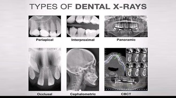
Another type of dental x-ray is the panoramic x-ray. This x-ray provides a broad view of the entire mouth, including the jawbone, teeth, and surrounding structures. It is particularly useful in evaluating impacted teeth, detecting cysts or tumors, and assessing the overall alignment of the teeth and jaw. With panoramic x-rays, dentists can make informed decisions regarding orthodontic treatment, oral surgery, and other necessary dental procedures.
In conclusion, understanding the different types of dental x-rays and their uses is critical in providing comprehensive oral healthcare. From detecting cavities to assessing jaw alignment, dental x-rays enable dentists to make accurate diagnoses and develop effective treatment plans. By utilizing these invaluable tools, dental professionals can enhance patient care and ensure optimal oral health outcomes.
Dental X-rays play a crucial role in early detection of oral health issues. These diagnostic images provide valuable insights for dentists, allowing them to identify and address problems before they escalate. By uncovering hidden conditions and abnormalities beneath the surface, dental X-rays enable dentists to provide timely treatment and preventive care to their patients.
One of the key benefits of dental X-rays is their ability to detect cavities at an early stage. While visual examinations can reveal obvious signs of tooth decay, X-rays can uncover hidden decay between teeth or beneath existing fillings. This allows dentists to address cavities before they cause significant damage, preserving the tooth structure and preventing more extensive treatments like root canals or extractions. Additionally, dental X-rays help in identifying bone loss, impacted teeth, and infections in the jaw, enabling dentists to provide appropriate interventions and prevent further complications.
Overall, the inclusion of dental X-rays in routine oral health care allows for early detection of issues that may not be visible during regular examinations. By enabling dentists to identify problems in their infancy, X-rays help promote proactive dental care and can save patients from experiencing more severe conditions down the line. Regular X-rays can be a valuable tool in maintaining optimal oral health and preventing long-term dental complications.
Dental X-rays play a critical role in diagnosing cavities and tooth decay that may not be visible to the naked eye. These highly effective diagnostic tools provide dentists with detailed images of the teeth and supporting structures, allowing for early detection and prevention of dental problems. By utilizing X-rays, dentists can identify cavities in their early stages, enabling prompt treatment and the preservation of natural tooth structure.
This is particularly crucial in pediatric dentistry, as children may not always be able to accurately communicate or recognize dental issues. With the help of dental X-rays, dentists can detect cavities in primary teeth, preventing potential complications and reducing the need for extensive treatments later on.
Moreover, dental X-rays can assist in detecting tooth decay that occurs between teeth or beneath existing fillings. These hard-to-reach areas often harbor hidden decay, which can progress rapidly if left untreated. X-rays allow dentists to visualize these areas, helping them identify and address decay before it leads to further damage or infection. By catching cavities and tooth decay early on through the use of dental X-rays, dentists can provide timely and targeted interventions, preserving the overall oral health and well-being of their patients.
Gum disease, also known as periodontal disease, is a common oral health condition that affects millions of people worldwide. It is characterized by inflammation of the gums, which can eventually lead to the destruction of the surrounding tissues and even tooth loss if left untreated. While regular dental exams and cleanings play a crucial role in maintaining gum health, dental X-rays are essential for assessing the extent of gum disease and developing appropriate treatment plans.
Table
| Aspect Assessed by Dental X-rays | Role in Gum Disease Evaluation |
|---|---|
| Amount of bone present | Helps determine bone loss due to periodontal disease. |
| Condition of the alveolar crests | Indicates the health of the supporting bone structure. |
| Bone loss in the furcation areas | Reveals the extent of bone loss around the roots of teeth. |
| Width of the PDL space | Abnormal width can suggest periodontal inflammation. |
| Local factors | Identifies calculus, poorly contoured restorations, and caries that can exacerbate gum disease. |
Dental X-rays provide a comprehensive view of the teeth, jawbone, and supporting structures that cannot be seen with the naked eye. In the case of gum disease, X-rays can reveal important information such as the presence of bone loss, which is a critical indicator of the severity of the condition. By capturing detailed images of the teeth and surrounding tissues, X-rays help dentists determine the extent of damage caused by gum disease and develop personalized treatment approaches. This could include deep cleaning procedures like scaling and root planing, as well as surgical interventions in more advanced cases. Ultimately, dental X-rays enable dentists to diagnose gum disease accurately and provide targeted care to improve oral health outcomes.
Orthodontic treatments play a vital role in correcting misaligned teeth, improving bite function, and enhancing a patient’s overall oral health and aesthetics. When it comes to planning these treatments, dental x-rays serve as a valuable tool for orthodontists in accurately assessing the position of the teeth and the underlying bone structure.
Dental x-rays aid orthodontic treatment planning by providing detailed images of the teeth, roots, and jawbone. These images allow orthodontists to evaluate the eruption pattern of the permanent teeth, determine the presence of any impacted teeth, and assess the alignment of the dental arches. By analyzing the x-ray images, orthodontists can identify any abnormalities, such as extra or missing teeth, and identify the most appropriate treatment approach for each individual patient.
Additionally, dental x-rays help orthodontists in assessing the health of the teeth and supporting structures before initiating orthodontic treatment. X-rays reveal any underlying dental issues that may need to be addressed before braces or aligners are applied. These could include the presence of cavities, gum disease, or root resorption. By addressing these issues early on, potential complications during orthodontic treatment can be minimized, leading to better treatment outcomes.
Overall, dental x-rays serve as an essential tool in the planning and execution of orthodontic treatments. They provide orthodontists with detailed and valuable information, allowing them to create individualized treatment plans and achieve optimal results for their patients. With the assistance of dental x-rays, orthodontic treatments can be planned more efficiently, taking into account the unique needs of each patient and resulting in healthier, straighter smiles.
One of the most common myths surrounding dental X-rays is that they are unsafe due to the exposure to radiation. However, it is important to debunk this myth as dental X-rays are, in fact, safe and pose minimal risk to patients. The amount of radiation emitted during a dental X-ray procedure is extremely low, equivalent to the radiation exposure that we receive from our natural environment on a daily basis. In fact, the American Dental Association states that the risk of developing any adverse effects from dental X-rays is extremely low, especially when compared to the benefits they provide in terms of early detection and diagnosis of oral health issues.
Table
| Myth | Fact |
|---|---|
| Dental X-rays pose a significant radiation risk. | Dental X-rays emit low levels of radiation and are considered safe with proper precautions. |
| There are no safety measures during dental X-rays. | Dental practices use lead aprons and thyroid collars to minimize exposure, and digital systems require less radiation than traditional methods. |
| Dental X-rays are done excessively frequently. | The frequency of dental X-rays is based on individual needs and risk factors, typically recommended every 1 to 2 years for most adults. |
Another myth that needs to be debunked is the belief that dental X-rays can cause cancer. This misconception stems from a misunderstanding of the radiation levels associated with dental X-rays. The truth is that the amount of radiation used in dental X-rays is so low that the risk of developing cancer from these procedures is negligible. In fact, studies have shown that the risk of developing cancer from dental X-rays is incredibly low, even for patients who require frequent X-rays for various dental treatments. It is important for patients to understand that the benefits of dental X-rays far outweigh the minimal risks associated with them.
Dental X-rays play a crucial role in the field of oral surgery, aiding dentists in diagnosing and treating various conditions. These diagnostic images provide valuable insights into oral health, allowing surgeons to thoroughly examine the underlying structures before performing any surgical procedures.
Before any oral surgery, dentists rely on dental X-rays to assess the current condition of the patient’s teeth, gums, and jawbone. These X-rays help identify the presence of any underlying dental issues, such as impacted teeth, root canal infections, or bone loss. By having a clear and comprehensive view of the oral structures, surgeons can develop precise treatment plans and make informed decisions during the surgical process.
During oral surgery, dental X-rays continue to assist in ensuring the success of the procedure. They guide surgeons in determining the exact placement of dental implants, identifying anatomical structures to avoid any potential complications. Moreover, X-rays enable dentists to monitor the progress of bone grafts or other regenerative treatments, allowing for timely adjustments and optimal outcomes.
With the aid of dental X-rays, oral surgeons can provide safer and more effective treatment options, enhancing the overall success of surgical procedures. These diagnostic images contribute to the planning, execution, and evaluation of oral surgeries, ensuring better patient outcomes and improved oral health.
Dental X-rays play a crucial role in identifying tumors and other abnormalities within the oral cavity. These imaging techniques provide dentists with a detailed view of the structures beneath the surface, helping them to detect certain conditions that may not be visible during a regular dental examination.
One of the most significant benefits of dental X-rays in detecting tumors is their ability to identify early-stage oral cancers. According to the American Cancer Society, the five-year survival rate for oral cancer is approximately 84% when detected early, compared to only 39% when the cancer has already spread to other parts of the body. Dental X-rays can help identify suspicious growths or abnormal changes in the tissues, allowing for timely diagnosis and intervention. Additionally, X-rays can also assist in detecting cysts, abscesses, and other abnormalities that may require further evaluation or treatment.
While the importance of dental X-rays in identifying tumors and other abnormalities cannot be overstated, it’s essential to note that these imaging techniques should be used judiciously, considering the potential risks associated with radiation exposure. Dentists follow strict guidelines and protocols to minimize radiation exposure and ensure patient safety. With advancements in technology, dental X-rays have become safer and more precise, allowing for accurate diagnosis and improved patient care.
Routine dental check-ups are an essential part of maintaining good oral health. One of the important components of these check-ups is dental x-rays. These x-rays provide valuable insights into the overall dental health of a person by detecting issues that may not be visible to the naked eye. However, it is crucial to determine the frequency of dental x-rays to minimize unnecessary exposure to radiation.
The frequency of dental x-rays varies depending on certain factors such as age, oral health status, and risk of developing dental problems. For adults with good oral health, it is generally recommended to have bitewing x-rays once every two to three years. On the other hand, children and teenagers are advised to have bitewing x-rays once a year to monitor the development of their teeth and identify any potential issues early on.
It is important to note that these guidelines may differ for individuals with higher risk factors. For example, individuals with a history of dental problems or those who are undergoing orthodontic treatment may require more frequent dental x-rays. Additionally, individuals who are experiencing symptoms such as tooth pain or swelling may need immediate x-rays to help diagnose and treat the underlying issue.
Overall, the frequency of dental x-rays should be determined on a case-by-case basis, taking into consideration the individual’s oral health status and specific risk factors. Dentists are trained to assess each patient’s needs and make informed decisions regarding the appropriate timing and frequency of dental x-rays to ensure that they benefit from these diagnostic tools while minimizing unnecessary radiation exposure. By following these guidelines, individuals can maintain their oral health and address any potential issues before they become more severe.
Exposure to radiation is a concern for both patients and dental professionals during dental x-ray procedures. While the risks of radiation from dental x-rays are minimal, it is important to take steps to minimize exposure and ensure the safety of patients.
One way to minimize radiation exposure is by using digital x-ray technology. Digital x-rays require significantly less radiation than traditional film-based x-rays, reducing the patient’s exposure by up to 90%. Additionally, digital x-rays offer better image quality, allowing for more accurate diagnoses. It is recommended for dental practices to invest in digital x-ray systems to provide the best possible care while minimizing radiation exposure.
Another way to minimize radiation exposure is by using protective barriers. Lead aprons and thyroid collars are commonly used to shield sensitive areas of the body from radiation. These protective barriers are designed to absorb and block radiation, providing an extra layer of safety during the x-ray procedure.
By employing digital x-ray technology and utilizing protective barriers, dental professionals can ensure the safety and well-being of their patients while still obtaining the necessary diagnostic information. It is important for dental practices to adhere to radiation safety protocols and stay updated on advancements in technology to continually improve the patient experience and reduce potential risks.
Preparing for Dental X-rays: What to Expect during the Procedure
When it comes to preparing for dental x-rays, it’s important to know what to expect during the procedure. Dental x-rays are a valuable tool in diagnosing and detecting oral health issues that may not be visible to the naked eye. Before the procedure begins, it’s essential to disclose any relevant medical history, such as pregnancy or previous radiation exposure, to ensure the safety and accuracy of the x-rays.
During the procedure, the dental professional will position a specialized x-ray machine near your mouth and ask you to bite down on a small piece of plastic or hold a sensor against your teeth. The x-ray machine will emit a small amount of radiation to capture images of your teeth and jawbone. While the process is quick and painless, the dental professional may need to take multiple x-rays from different angles to gather a comprehensive view of your oral health. By knowing what to expect during the procedure, you can ensure a smooth and efficient experience while reaping the benefits of this important diagnostic tool.
Interpreting dental X-ray images is a crucial part of accurate diagnosis in dentistry. Dentists rely on these images to identify and understand various oral health issues that may not be visible to the naked eye. By analyzing the details captured in X-ray images, dentists can gain valuable insights into potential problems, allowing for timely intervention and appropriate treatment.
When dentists interpret dental X-ray images, they carefully examine the different structures of the teeth, gums, and jawbone. They look for signs of tooth decay, cavities, gum disease, and abnormalities such as tumors or cysts. By studying the size, shape, and density of these structures, dentists can evaluate the health of the teeth and surrounding tissues.
Additionally, dentists use dental X-rays to assess the alignment of teeth and plan orthodontic treatments. These images provide crucial information about the spacing between teeth, eruption patterns, and any potential issues that may require intervention. By analyzing X-ray images, dentists can determine the most effective course of action to achieve optimal oral health and a beautiful smile.
Overall, the interpretation of dental X-ray images plays a vital role in accurate diagnosis in dentistry. Dentists utilize their expertise to analyze these images, identify potential issues, and develop personalized treatment plans. With the help of dental X-rays, dentists can provide patients with the highest level of care, ensuring long-lasting oral health and well-being.
Advancements in imaging technology have revolutionized the field of dental x-rays, paving the way for a promising future in oral healthcare. With the development of digital radiography, traditional film-based x-rays are being gradually replaced, offering numerous advantages for both dentists and patients.
Digital radiography eliminates the need for chemical processing and provides instant results, allowing dentists to quickly and accurately diagnose dental conditions. Moreover, the images can be easily shared and stored electronically, reducing the risk of damage or loss. The clarity and resolution of digital images are also superior, enabling dentists to detect even the smallest abnormalities with greater precision. Additionally, digital x-rays significantly reduce radiation exposure, making them a safer option for patients of all ages. This groundbreaking technology not only enhances the efficiency and accuracy of dental diagnostics but also contributes to overall patient satisfaction and improved oral health outcomes.
As technology continues to evolve, the future of dental x-rays holds even more promising advancements. One such innovation is the cone beam computed tomography (CBCT), which captures 3D images of the oral structures. This powerful tool provides dentists with a comprehensive view of the teeth, bones, nerves, and soft tissues, facilitating more precise diagnosis and treatment planning. CBCT imaging is particularly valuable in complex cases such as orthodontics, implant placement, and dental surgeries. With its ability to accurately assess anatomical structures, CBCT offers an invaluable resource for enhancing procedural success and optimizing patient care.
The future of dental x-rays is undeniably bright, with continued advancements in imaging technology revolutionizing the field of oral healthcare. As digital radiography becomes the standard, dentists can expect increased efficiency, improved accuracy, and enhanced patient safety. Furthermore, the emergence of 3D imaging like CBCT holds great promise in revolutionizing treatment planning and outcomes. With these advancements, dental professionals are well-equipped to provide the best possible care for their patients, ensuring optimal oral health for years to come.
Yes, dental x-rays are safe. The amount of radiation exposure from dental x-rays is minimal and well within acceptable limits. Dentists take necessary precautions to minimize radiation exposure, such as using lead aprons and collars.
The frequency of dental x-rays depends on your oral health condition and risk factors. As a general guideline, most adults require dental x-rays every 1-2 years, whereas children may need them more frequently. Your dentist will determine the appropriate interval based on your specific needs.
Yes, dental x-rays play a crucial role in diagnosing cavities. They can detect early signs of tooth decay that are not visible to the naked eye. This allows dentists to address cavities in their early stages, preventing further damage to the teeth.
Dental x-rays provide valuable information about the position and alignment of teeth, jawbone structure, and other factors that influence orthodontic treatment. Orthodontists use these images to create effective treatment plans and determine the appropriate course of action.
Yes, dental x-rays can help identify tumors, cysts, and other abnormalities in the oral and maxillofacial region. They provide dentists with a clear view of the underlying structures, allowing for early detection and timely intervention.
During a dental x-ray procedure, you will be asked to wear a lead apron to protect the rest of your body from radiation. The dentist or dental technician will position a small x-ray sensor or film inside your mouth and take the necessary images. The process is quick and painless.
Dentists are trained to interpret dental x-ray images accurately. They look for signs of cavities, bone loss, gum disease, tumors, and other abnormalities. The interpretation of these images helps dentists make an accurate diagnosis and develop an appropriate treatment plan.
The future of dental x-rays involves advancements in imaging technology, such as digital x-rays and cone beam computed tomography (CBCT). Digital x-rays offer higher image quality, reduced radiation exposure, and instant results. CBCT provides 3D images, allowing for more precise diagnostics and treatment planning.