Physical Address
304 North Cardinal St.
Dorchester Center, MA 02124
Physical Address
304 North Cardinal St.
Dorchester Center, MA 02124

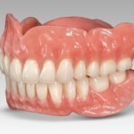
Explore the benefits of digital radiography for accurate dental diagnosis and treatment.
The field of dental imaging technology has come a long way in its evolution, revolutionizing the way dental professionals capture and analyze images of the oral cavity. From the introduction of traditional X-rays to the more recent advancements in digital radiography, the evolution of dental imaging technology has greatly improved the accuracy and efficiency of diagnosing oral health conditions.
Traditional dental X-rays were a significant breakthrough in the early days of dental imaging. However, they had their limitations, including the need for film processing and the exposure of patients to higher levels of radiation. With the advent of digital radiography, these limitations have been overcome. Digital radiography offers numerous advantages, including instant image capture, reduced radiation exposure, and the ability to enhance and manipulate images for better analysis. This technology has not only facilitated more accurate and reliable diagnoses but has also improved the overall patient experience by minimizing discomfort and limiting the need for retakes due to overexposure or underexposure.
As dental professionals continue to explore and embrace the benefits of digital radiography, it is anticipated that further advancements and innovations will shape the future of dental imaging technology. These advancements may include the integration of artificial intelligence for computer-aided diagnosis, the development of smaller and more portable imaging devices, and the utilization of 3D imaging techniques for enhanced visualization. The evolution of dental imaging technology is an ongoing process, driven by the ever-growing need for more efficient and precise methods of diagnosing and treating oral health conditions.
Digital radiography is a vital component of modern dental practices, offering numerous benefits over traditional X-ray methods. By utilizing digital sensors instead of film, dentists can capture high-resolution images of a patient’s teeth and oral structures with greater ease and accuracy. These images are then instantly available for analysis, diagnosis, and treatment planning, ensuring efficient and effective care.
One of the key advantages of digital radiography is its ability to reduce radiation exposure for both patients and dental professionals. Compared to traditional X-rays, digital radiography requires significantly less radiation to produce diagnostic images. This not only enhances patient safety but also contributes to a more sustainable and environmentally friendly approach to dental imaging. Additionally, digital radiography eliminates the need for film processing, saving time and resources while promoting a paperless dental practice environment.

Overall, understanding the basics of digital radiography is crucial for dental professionals seeking to provide the best possible care to their patients. By embracing this innovative technology, dentists can enhance diagnostic accuracy, improve treatment planning, and streamline workflow efficiency for better patient outcomes. As digital radiography continues to evolve and integrate with advanced software and systems, the future of dental imaging looks promising, promising even greater advancements in this essential aspect of oral healthcare.
Digital radiography has revolutionized the field of dental imaging, offering numerous advantages over traditional X-rays. One significant advantage is the reduced exposure to radiation for both patients and dental professionals. Digital radiography uses up to 90% less radiation than traditional X-rays, ensuring the safety and well-being of individuals undergoing dental procedures.
Moreover, digital radiography provides superior image quality, allowing for more accurate and detailed diagnosis. The images captured through digital radiography are instantly available, eliminating the need for film development and reducing processing time. This not only saves time but also allows dentists to enhance the image quality through various digital tools and techniques. With clearer and more precise images, dentists can detect dental issues at an early stage and provide prompt and effective treatment, resulting in improved patient outcomes.
| Advantages of Digital Radiography over Traditional X-rays |
|---|
| Faster Image Acquisition |
| Higher Image Quality |
| Lower Radiation Exposure |
| Enhanced Image Manipulation |
| Digital Storage and Retrieval |
| Integration with Electronic Health Records (EHR) |
| Reduced Environmental Impact |
| Improved Workflow |
| Telemedicine Capabilities |
Digital radiography has revolutionized the field of dentistry by enhancing accuracy in dental diagnosis. Traditional X-ray films have long been used to capture images of the teeth and jaws, but they often provide limited information and require longer processing times. With digital radiography, dentists can now obtain high-resolution images within seconds, allowing for a more efficient and precise assessment of a patient’s oral health.
One key advantage of digital radiography is its ability to provide detailed images that can be magnified, zoomed in, and manipulated to reveal even the smallest dental problems. Dentists can analyze these images on a computer screen, adjusting contrast and brightness levels to better visualize teeth, bones, and soft tissues. This level of customization significantly enhances accuracy in dental diagnosis, enabling dentists to identify issues such as cavities, bone loss, impacted teeth, and abnormalities in the jaw or sinus areas. Moreover, digital radiography enables dentists to compare images taken at different time points, aiding in the detection and monitoring of dental conditions over time.
In addition to improved diagnostic capabilities, digital radiography also reduces radiation exposure for patients. Unlike traditional X-rays that require higher levels of radiation to create an image, digital radiography uses a sensor that is highly sensitive to X-rays and requires significantly less radiation. This reduction in radiation exposure not only promotes patient safety but also makes digital radiography a preferred choice, particularly for vulnerable populations such as children and pregnant women. By enhancing accuracy and minimizing radiation risks, digital radiography has become an indispensable tool in modern dental practices, enabling dentists to provide more precise and personalized treatment plans for their patients.
Digital radiography has revolutionized the field of dental imaging, offering dentists a range of options to capture high-quality images of the oral cavity. There are several different types of digital radiography systems available, each with its own unique advantages and applications.
One popular type of digital radiography system is the intraoral sensor. This small, flat device is inserted into the patient’s mouth to capture detailed images of individual teeth. Intraoral sensors offer excellent image resolution and can be used to detect cavities, monitor the progression of dental diseases, and aid in treatment planning. They are particularly useful in pediatric dentistry, where small mouths can make traditional x-ray film placement difficult.
Another type of digital radiography system is the panoramic unit, which produces a wide-angle view of the entire oral cavity. This type of imaging is useful for evaluating the overall health of the teeth, jawbone, and surrounding structures. Panoramic radiographs are commonly used for orthodontic evaluation, wisdom teeth assessment, and detecting pathology in the maxillofacial region.
| Type of Digital Radiography System | Description |
|---|---|
| Computed Radiography (CR) | Uses a photostimulable phosphor plate to capture X-ray images. The plate is processed in a reader which extracts the image data digitally. CR systems are versatile and can be used with existing X-ray machines. |
| Direct Radiography (DR) | Utilizes a digital detector panel to directly capture X-ray images. There are two types: flat-panel detectors (FPD) and indirect-conversion detectors (ICD). DR systems offer faster image acquisition and higher image quality compared to CR. |
| Portable Digital Radiography | Designed for mobile use, these systems typically use either CR or DR technology. Portable systems are convenient for bedside imaging in hospitals, emergency situations, or in field environments. |
| Cone Beam Computed Tomography (CBCT) | Provides three-dimensional images by rotating an X-ray source and detector around the patient. CBCT is commonly used in dental and orthopedic applications for detailed imaging of structures such as teeth, bones, and joints. |
| Digital Mammography | Specifically designed for breast imaging, digital mammography systems use DR technology optimized for low-dose imaging of breast tissue. Digital mammography offers improved image quality and allows for computer-aided detection of abnormalities. |
| Fluoroscopy | Real-time imaging technique used for dynamic studies such as barium swallow, cardiac catheterization, and gastrointestinal exams. Digital fluoroscopy systems offer enhanced image quality, reduced radiation dose, and the ability to store and manipulate images digitally. |
Cone beam computed tomography (CBCT) is a more advanced type of digital radiography system that provides three-dimensional images of the oral and maxillofacial region. CBCT scans are invaluable in complex cases such as dental implant planning, orthodontic treatment, and diagnosing temporomandibular joint disorders. The ability to visualize anatomical structures in three dimensions allows for greater accuracy in treatment planning and surgical procedures.
In conclusion, exploring different types of digital radiography systems provides dentists with a range of imaging options to suit their specific needs. Whether using intraoral sensors, panoramic units, or CBCT scanners, dental professionals can now capture high-quality images of the oral cavity to aid in diagnosis, treatment planning, and monitoring oral health. The advancements in digital radiography technology have undoubtedly enhanced the accuracy and precision of dental imaging, ultimately leading to better patient care and outcomes.
Digital radiography equipment has revolutionized the field of dentistry, offering numerous key features and components that enhance the imaging process. One of the most notable features of digital radiography equipment is its ability to provide instant image results. Unlike traditional film-based X-rays, digital radiography saves time by eliminating the need for manual development and processing. Dentists and dental technicians can now view high-quality images within seconds, allowing for immediate diagnosis and treatment planning.
Another essential component of digital radiography equipment is the digital sensor. This small device captures the X-ray image and converts it into a digital format that can be viewed on a computer screen. Digital sensors are available in different sizes and shapes to accommodate various dental imaging needs, such as intraoral or extraoral imaging. Their high sensitivity and resolution ensure detailed and accurate images, providing dentists with a comprehensive view of the patient’s oral health.
In addition to instant results and advanced sensors, digital radiography equipment often includes user-friendly software. This software enables dentists to enhance, analyze, and manipulate the digital images, helping them to identify even the smallest dental issues. Software features may include measurement tools, image editing options, and the ability to compare previous and current images for tracking changes in the patient’s oral health.
Overall, the key features and components of digital radiography equipment contribute to improved efficiency and accuracy in dental imaging. With instant image results, high-quality sensors, and advanced software, dentists can diagnose conditions more quickly and provide targeted treatments, ultimately enhancing patient care.
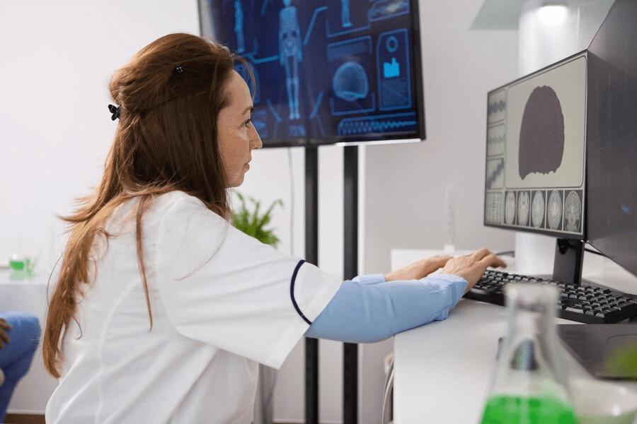
With advancements in digital radiography technology, software plays a vital role in the analysis of dental images. The software used in digital radiography analysis allows for the enhancement, manipulation, and examination of dental images, providing dentists with valuable information for diagnosis and treatment planning.
One of the key functions of software in digital radiography analysis is image optimization. By adjusting contrast, brightness, and sharpness, the software helps to improve the visibility of dental structures, enabling dentists to identify and assess dental conditions more accurately. Additionally, software allows for measurements to be taken directly on the digital images, providing precise data for treatment planning and monitoring progress over time. This level of detail and precision can significantly enhance the accuracy of dental diagnosis and aid in providing tailored treatment plans for patients.
| Role of Software in Digital Radiography Analysis |
|---|
| 1. Image Enhancement |
| Software allows for the enhancement of digital radiographs, improving clarity and contrast for better interpretation by radiologists. It includes tools for adjusting brightness, contrast, and sharpness. |
| 2. Measurement and Annotation |
| Software enables precise measurement of anatomical structures or lesions directly on digital images. Additionally, radiologists can annotate images with notes or arrows to highlight specific areas of interest. |
| 3. 3D Reconstruction |
| Advanced software provides capabilities for reconstructing 3D images from 2D radiographic data, allowing for better visualization of complex anatomical structures and pathology. |
| 4. Image Stitching |
| In cases where a single radiographic image does not capture the entire region of interest, software can stitch together multiple images to create a panoramic view, aiding in comprehensive analysis. |
| 5. Integration with PACS |
| Software seamlessly integrates with Picture Archiving and Communication Systems (PACS), enabling efficient storage, retrieval, and sharing of digital radiographic images across healthcare facilities. |
| 6. Computer-Aided Detection (CAD) |
| CAD software assists radiologists in detecting abnormalities or potential pathology by automatically identifying suspicious areas on radiographic images, helping to improve diagnostic accuracy. |
| 7. Quantitative Analysis |
| Software facilitates quantitative analysis of radiographic data, such as bone mineral density measurements or tumor volume calculations, providing objective metrics for assessment and monitoring. |
| 8. Workflow Optimization |
| By automating routine tasks, such as image processing and report generation, software streamlines the radiography analysis workflow, allowing radiologists to focus more on interpretation and diagnosis. |
| 9. Telemedicine and Teleradiology |
| Software enables remote access to digital radiographic images, facilitating telemedicine consultations and teleradiology services, particularly in underserved or remote areas. |
| 10. Quality Assurance |
| Software includes tools for quality assurance and compliance with regulatory standards, ensuring that digital radiography systems produce accurate and reliable results. |
Patient safety and radiation protection are paramount considerations in digital radiography. With advancements in technology, digital radiography has significantly reduced radiation exposure compared to traditional x-rays. The use of digital sensors and image receptors has not only improved image quality but also minimized the amount of radiation patients are exposed to during dental procedures.
To ensure patient safety, dental professionals should adhere to established guidelines and protocols for radiation protection. This includes the use of lead aprons, thyroid collars, and other protective equipment to shield patients from unnecessary radiation. Additionally, proper positioning and technique during image capture can help minimize retakes, reducing the overall amount of radiation exposure. Regular calibration and maintenance of digital radiography equipment also play a vital role in ensuring accurate and precise imaging, further enhancing patient safety and diagnostic reliability.
Digital radiography has revolutionized the field of dentistry, offering numerous advantages over traditional x-ray techniques. However, like any emerging technology, its implementation in dental practices can come with its fair share of challenges. Overcoming these challenges is crucial to ensure a seamless integration of digital radiography into the workflow of dental professionals.
One of the common challenges faced during the implementation of digital radiography is the initial cost involved. Upgrading to digital radiography equipment can be a significant investment for dental practices, particularly for smaller clinics with limited resources. However, it is important to consider the long-term benefits and cost savings that digital radiography offers. By eliminating the need for film, chemical developers, and storage space, digital radiography can result in substantial cost reductions in the long run. Additionally, many manufacturers offer financing options and leasing programs to make the transition more affordable for dental practices.
Another challenge in digital radiography implementation is the learning curve associated with mastering new software and equipment. Dental professionals may need to dedicate time and effort to become familiar with the features and functionalities of their chosen digital radiography system. This may require training sessions or workshops, which can be time-consuming. However, the investment in training is well worth it, as mastering the software and equipment will ultimately enhance accuracy in dental diagnosis and streamline the workflow in the long run.
In conclusion, while there may be challenges in implementing digital radiography in dental practices, it is important for dental professionals to embrace this technology to stay ahead in the field. The initial costs can be justified by the long-term financial benefits, and the learning curve can be overcome through dedicated training. By overcoming these challenges, dental professionals can unlock the full potential of digital radiography and provide superior care to their patients.
When it comes to capturing high-quality digital dental images, following best practices is essential. Accurate and clear images are crucial for effective diagnosis, treatment planning, and case presentation. Here are some key strategies to ensure that you achieve optimal results with your digital radiography equipment:
1. Proper positioning: Correct patient positioning is critical for obtaining accurate dental images. Make sure to instruct the patient on how to position their head, maintain the correct angle, and bite down properly on the sensor or film. Additionally, ensure that the sensor or film is aligned correctly within the patient’s mouth to capture the desired area.
2. Exposure settings: Adjusting the exposure settings appropriately is crucial to achieving clear and detailed images. Take into account factors such as patient age, anatomy, and the specific dental region being imaged. Consider using exposure assistance devices or software features that can help guide you in selecting the optimal exposure settings.
By implementing these best practices, dental professionals can increase the likelihood of capturing high-quality digital dental images, enhancing their ability to provide accurate diagnoses and deliver effective treatment plans. Stay tuned for more insights on how digital radiography is transforming the field of dentistry and improving patient care.
Proper image storage and management play a crucial role in digital radiography, ensuring that patient records are organized, accessible, and secure. With the advancement of technology, dental practices have shifted from traditional physical film to digital images, which offer numerous benefits such as easier storage, faster retrieval, and enhanced image quality. However, without an efficient system in place for managing and storing these digital images, valuable patient data may be at risk of being lost, compromised, or even misinterpreted.
One of the key advantages of digital radiography is that it allows for the seamless integration of patient records into a dental practice’s computer system. This enables dental professionals to easily access and review a patient’s previous radiographic images, which is particularly crucial for treatment planning and case presentations. Through proper image storage and management, dental practices can maintain a comprehensive and up-to-date record of their patients’ dental health, facilitating more accurate diagnoses, effective treatment plans, and improved continuity of care. Additionally, a well-organized digital image system allows for efficient collaboration between dental professionals, providing a platform for radiographic image sharing and consultation, contributing to enhanced communication and better patient outcomes.
In conclusion, the importance of proper image storage and management in digital radiography cannot be understated. It not only ensures the integrity and accessibility of patient records but also promotes efficient communication and collaboration within a dental practice. By adopting best practices for image storage and management, dental professionals can harness the full potential of digital radiography, ultimately enhancing their ability to provide quality dental care to their patients.
Integrating digital radiography into the dental practice workflow has revolutionized the way dental professionals diagnose and treat patients. With its numerous benefits and advancements, digital radiography has become an essential tool in modern dentistry.
One of the key advantages of digital radiography is its ability to provide immediate results. Unlike traditional X-rays, which required film development, digital radiography allows dentists to view high-quality images within seconds. This not only saves time for both the dental team and the patient, but it also enables dentists to make real-time diagnoses and provide prompt treatment recommendations. Additionally, the digital format of these images allows for easy storage, retrieval, and sharing, enhancing communication and collaboration among dental professionals. With the integration of digital radiography into the dental practice workflow, dental teams can streamline their processes, improve efficiency, and ultimately deliver better patient care.
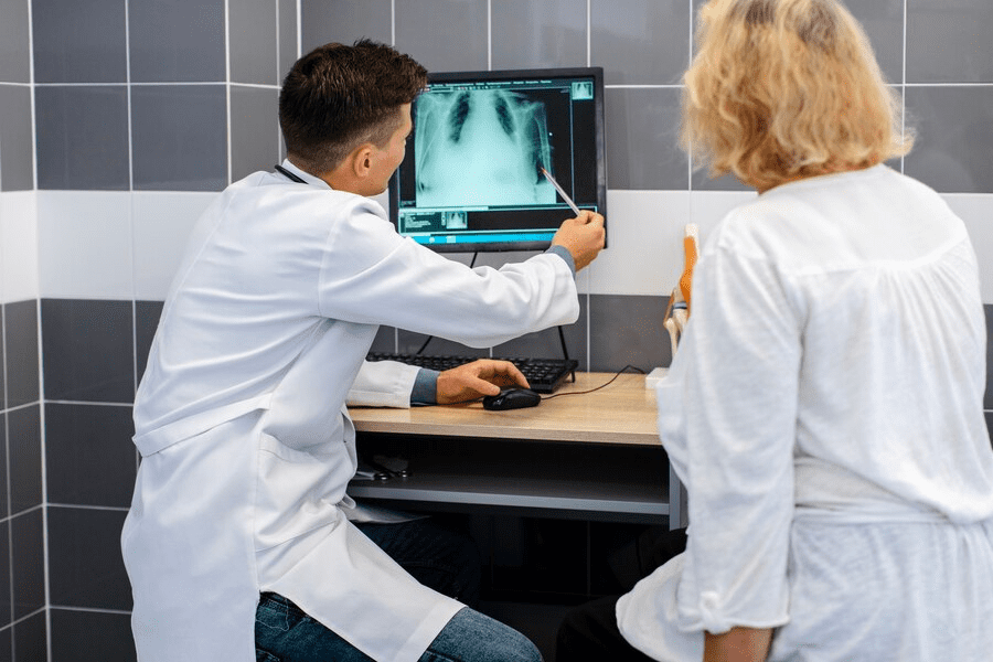
Effective communication and collaboration are vital in the dental field, as they play a crucial role in ensuring optimal patient care. With the advent of digital radiography, the way we communicate and collaborate as dental professionals has been revolutionized. Digital radiography allows for instant access to high-quality images, which can be easily shared and discussed among dental team members and with patients themselves.
One of the key advantages of digital radiography in enhancing communication is the ability to quickly and accurately diagnose and discuss treatment options with patients. By displaying the digital radiographs on chairside monitors or computer screens, dentists can easily explain and illustrate dental conditions to their patients. This visual aid not only helps in patient education, but also builds trust and confidence as patients are actively involved in the treatment planning process. Additionally, digital radiography enables seamless sharing of images with specialists and other healthcare professionals, facilitating collaborative approaches to complex cases.
Moreover, digital radiography results can be conveniently stored in electronic patient records, creating a centralized database that can be accessed by the entire dental team. This promotes efficient communication and collaboration, as team members can review and analyze the same images, leading to better coordination of treatment plans. Furthermore, digital radiography systems often have features that allow for annotations and measurements to be added directly onto the images, facilitating more precise communication among dental professionals and enhancing interdisciplinary collaboration.
In summary, the integration of digital radiography into dental practices has undoubtedly enhanced communication and collaboration among dental professionals and with patients. The ability to instantly view, discuss, and share high-quality images has transformed the way dental teams work together, leading to improved patient care and outcomes. Digital radiography has revolutionized communication in the dental field, paving the way for more effective collaboration in treatment planning and case management.
Digital radiography has revolutionized the way dental professionals approach treatment planning and case presentation. With its advanced imaging capabilities, digital radiography provides a clear and detailed view of a patient’s dental structures, enabling dentists to accurately diagnose and develop comprehensive treatment plans. This technology has a significant impact on improving patient outcomes and enhancing the overall dental experience.
One of the key benefits of digital radiography in treatment planning is its ability to facilitate precise and accurate measurements. Traditional x-rays often suffer from distortion and magnification, making it challenging to obtain precise measurements for treatment planning. However, digital radiography eliminates these issues by providing highly detailed and distortion-free images. Dentists can use these images to assess the dimensions of the teeth, bone structure, and soft tissues accurately. This precise mapping allows for more accurate treatment planning and enables dentists to develop personalized treatment approaches tailored to their patients’ specific needs.
Moreover, digital radiography allows for better case presentation to patients. With the help of high-resolution images, dentists can visually convey the dental issues and treatment options to patients in a clear and comprehensive manner. This visual aid plays a crucial role in patient education and helps patients understand the severity of their condition and the potential treatment outcomes. By having a visual representation of their dental health, patients are more likely to make informed decisions and actively participate in their treatment journey.
In conclusion (The Conclusion section will be written separately), digital radiography has a profound impact on treatment planning and case presentation. Its advanced imaging capabilities provide dentists with accurate measurements and enable them to visually communicate with patients effectively. The integration of digital radiography into the dental practice workflow has enhanced the overall dental experience, leading to improved patient outcomes and treatment success.
Digital radiography has become increasingly popular in dental practices due to its cost-effectiveness compared to traditional X-ray systems. The initial investment in digital radiography equipment may be higher, but the long-term benefits outweigh the costs. With digital radiography, there is no need for film development, chemicals, and storage space, which significantly reduces the expenses associated with traditional X-rays. Furthermore, digital radiography allows for image enhancement and manipulation, eliminating the need for retakes and further reducing costs.
Studies have demonstrated the cost-effectiveness of digital radiography in dental practices. One study conducted by the University of California, San Francisco found that the use of digital radiography resulted in a 42% reduction in per-patient costs compared to traditional X-rays. The study also reported a reduction in the number of retakes and environmental benefits associated with the elimination of film and chemical waste.
In addition to cost savings, digital radiography offers improved efficiency and workflow in dental practices. The instant image capture and ability to transfer images electronically allows for faster diagnoses and treatment planning. This can lead to enhanced patient satisfaction and increased productivity for the dental team.
As dental practices continue to embrace digital technology, the cost-effectiveness of digital radiography is becoming increasingly evident. By making the initial investment in digital radiography equipment, dental practices can experience long-term financial savings, improved workflow, and enhanced patient care.
Emerging technologies and innovations are set to revolutionize the field of digital radiography, offering exciting possibilities for dental practices and enhancing patient care. One such advancement is the development of 3D imaging techniques, which provide a more comprehensive view of the oral cavity and facilitate precise diagnosis and treatment planning. With the ability to visualize anatomical structures in three dimensions, dentists can better assess the extent of dental conditions such as impacted teeth, fractures, or cysts. This breakthrough technology allows for improved accuracy in treatment, resulting in optimal outcomes for patients.
Another area of innovation in digital radiography is the integration of artificial intelligence (AI) algorithms. AI has the potential to automate and expedite the analysis of dental images, reducing the time required for interpretation and increasing efficiency in dental practices. By training algorithms on vast datasets, AI can assist in detecting and diagnosing dental abnormalities, leading to early intervention and improved prognosis. Additionally, AI can help in the prediction of treatment outcomes and aid in the development of personalized treatment plans.
Digital radiography is an advanced imaging technology that allows dental professionals to capture and store dental X-ray images electronically.
Digital radiography offers several advantages, including reduced radiation exposure, faster image acquisition, improved image quality, and the ability to enhance and manipulate images for better diagnosis.
There are several types of digital radiography systems, including intraoral sensors, phosphor plate systems, and cone beam computed tomography (CBCT) scanners.
Digital radiography allows dental professionals to enhance and manipulate images, leading to improved accuracy in diagnosing dental conditions such as cavities, bone loss, and abnormalities.
When selecting digital radiography equipment, it is important to consider factors such as image resolution, sensor size, software compatibility, ease of use, and durability.
Software is an essential component of digital radiography systems as it allows for image viewing, enhancement, manipulation, measurement, and storage, enabling efficient analysis and diagnosis.
Dental professionals should follow proper radiation safety protocols, such as using lead aprons and thyroid collars, limiting exposure time, and regularly calibrating and maintaining the equipment.
Some common challenges in implementing digital radiography include initial costs, staff training, transitioning from traditional X-rays, and ensuring compatibility with existing practice management systems.
To capture high-quality images, it is important to ensure proper positioning of the patient, use appropriate exposure settings, maintain equipment cleanliness, and regularly calibrate the sensors.
Digital dental images should be stored securely in a protected electronic format and backed up regularly. Implementing a robust data management system is crucial to ensure easy access and retrieval.
To integrate digital radiography seamlessly, dental practices should create a standardized process for image acquisition, analysis, sharing with colleagues or specialists, and incorporating results into treatment plans.
Digital radiography provides detailed and accurate images that aid in treatment planning, allowing dental professionals to communicate and present treatment options to patients more effectively.
While the initial investment in digital radiography equipment can be significant, the long-term cost savings, improved efficiency, and enhanced diagnostic capabilities make it a cost-effective solution for dental practices.
The future of digital radiography holds promise for advancements such as artificial intelligence for automated diagnosis, improved image resolution, faster image processing, and integration with other digital technologies in dentistry.