Physical Address
304 North Cardinal St.
Dorchester Center, MA 02124
Physical Address
304 North Cardinal St.
Dorchester Center, MA 02124

Understand how dentists protect you from radiation during X-rays.
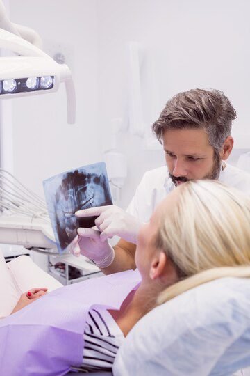
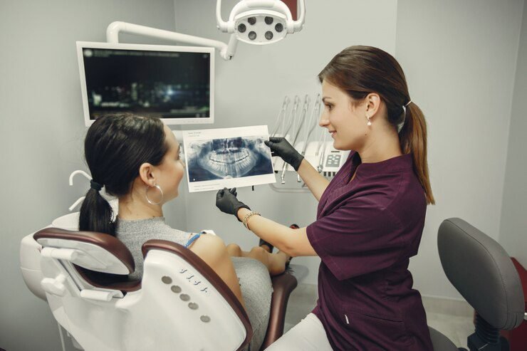
Dental X-rays play a crucial role in the diagnosis of oral health issues. They provide valuable information that cannot be obtained through a visual examination alone. X-rays allow dentists to see beneath the surface of the teeth and gums, helping them detect hidden problems such as cavities, infections, bone loss, and impacted teeth. This early detection enables dentists to develop appropriate treatment plans and prevent further damage to the teeth and surrounding tissues.
Furthermore, X-rays are especially important for pediatric dentistry. Children may not always be able to express their dental concerns or cooperate fully during examinations. X-rays help dentists identify any underlying problems that are not yet visible, ensuring that children receive the necessary treatment to maintain their oral health. Additionally, X-rays aid in monitoring the growth and development of children’s teeth, allowing dentists to anticipate any potential issues and intervene at the appropriate time. Overall, the use of X-rays in dental diagnosis is indispensable for providing accurate and comprehensive care to patients of all ages.

Dental X-rays play a crucial role in dental diagnosis, allowing dentists to identify oral health issues that may not be visible to the naked eye. There are several types of dental X-rays that are commonly used, each serving a specific purpose in dental imaging.
One common type of dental X-ray is the bitewing X-ray. This type of X-ray provides a detailed view of the upper and lower teeth in a specific area of the mouth. It is commonly used to detect cavities between the teeth, as well as to assess the health of the supporting bone structure.
Another type of dental X-ray is the periapical X-ray. This X-ray focuses on a single tooth, capturing images of the entire tooth, from the crown to the tip of the root. Periapical X-rays are useful for identifying issues such as abscesses, cysts, and other abnormalities that may be affecting a specific tooth.
Dental x-rays are an essential tool for dentists in detecting and diagnosing oral health issues. These diagnostic images provide valuable insights that cannot be obtained through visual examination alone. By capturing detailed images of the teeth, gums, and surrounding structures, dental x-rays enable dentists to identify and address a wide range of dental problems.
One of the key benefits of dental x-rays is their ability to reveal hidden dental issues. X-rays can detect tooth decay in its early stages, even before symptoms become apparent. This allows dentists to intervene and prevent the decay from progressing further. In addition, dental x-rays can identify infections in the root canal, detect cysts or tumors in the jawbone, and evaluate the status of developing teeth in children. By providing a comprehensive view of the oral structures, dental x-rays aid dentists in formulating accurate diagnoses and creating personalized treatment plans.
Moreover, dental x-rays are particularly valuable in monitoring the progression of dental diseases over time. By comparing previous x-rays with current ones, dentists can track the development of cavities, gum disease, and bone loss. This enables them to intervene at the right time and prevent further deterioration. For instance, x-rays can help dentists assess the effectiveness of orthodontic treatment, ensuring that the teeth are moving into their proper positions. Regular dental x-rays, combined with thorough clinical examinations, play a crucial role in maintaining optimal oral health and preventing potential complications.
X-rays have long been a vital tool in the field of dentistry, providing valuable diagnostic information that helps identify and address oral health issues. However, there is a common misconception that exposure to X-rays can significantly increase the risk of cancer. This is simply not true.
Numerous scientific studies have shown that the low levels of radiation used in dental X-rays pose minimal risk to patients. In fact, the amount of radiation received during a dental X-ray is roughly equivalent to the natural background radiation we are exposed to on a daily basis. The American Dental Association (ADA) and other reputable organizations recognize the safety and importance of dental X-rays in enhancing patient care.
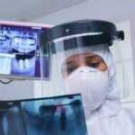
It’s important to note that dentists take several precautions to minimize radiation exposure during X-rays. Lead aprons and thyroid collars are routinely used to shield sensitive organs from any potential radiation. Additionally, dental X-ray rooms are equipped with proper shielding to ensure that radiation is contained within the space and does not pose a risk to staff or other patients.
In conclusion, the notion that X-rays used in dentistry pose a significant cancer risk is a myth that has been debunked by scientific evidence. The benefits of dental X-rays in detecting and addressing oral health issues far outweigh any minimal risk of radiation exposure. Dentists continue to follow strict safety protocols to ensure patient comfort and safety during X-ray procedures.
Dental X-rays are a valuable diagnostic tool in dentistry, providing crucial insights into oral health conditions that may not be visible to the naked eye. One common concern among patients is the amount of radiation exposure associated with dental X-rays. However, it is important to note that the levels of radiation emitted by dental X-rays are extremely low.
The radiation dose from dental X-rays is significantly lower compared to other medical imaging procedures, such as CT scans or chest X-rays. In fact, a dental X-ray exposes a patient to a radiation dose that is equivalent to the amount of radiation received from natural background sources in just a few days. To put it into perspective, the radiation dosage from a typical dental X-ray is approximately the same as one would receive from a cross-country airplane flight.
Dentists prioritize the safety and well-being of their patients, which is why they take stringent measures to protect individuals from radiation exposure during dental X-rays. One vital safety measure is the use of lead aprons and thyroid collars. These protective garments are designed to shield sensitive areas of the body, such as the reproductive organs and the thyroid gland, from radiation. By placing lead aprons over patients’ torsos and thyroid collars around their necks, dentists significantly reduce the risk of radiation reaching these critical areas.
Additionally, dental professionals employ proper shielding techniques in their X-ray rooms. These shielding measures include lead-lined walls, floors, and ceilings, which serve as barriers to prevent radiation from entering or leaving the X-ray room. Shielding not only protects patients but also ensures the safety of dental staff and other individuals in the vicinity. Dentists adhere to strict building codes and guidelines to construct X-ray rooms that meet or exceed the required level of radiation protection. These efforts ensure that patients undergo X-rays in a controlled and secure environment, minimizing their exposure to radiation.
Lead aprons and thyroid collars are essential protective measures used during dental x-rays to minimize radiation exposure to sensitive areas of the body. These protective devices are designed to shield the patient’s vital organs and thyroid gland from the small amount of radiation produced during the procedure.
Lead aprons are made of a heavy, radiation-resistant material that effectively blocks the penetration of x-rays. They are worn by patients to provide a barrier between the x-ray beam and their body. The aprons are strategically designed to cover the front of the body, extending from the neck to the knees, ensuring that the vital organs in the abdomen and pelvic region are shielded from radiation.
Thyroid collars, on the other hand, specifically aim to protect the thyroid gland from radiation exposure. The thyroid gland is a small, butterfly-shaped gland located in the front of the neck, and it is particularly sensitive to radiation. By placing a lead thyroid collar around the patient’s neck, dentists can further reduce the risk of radiation exposure to this delicate gland.
Together, lead aprons and thyroid collars work hand in hand to provide an additional layer of protection during dental x-rays. Dentists take great care in ensuring that these protective devices are properly positioned and securely fastened to maximize their effectiveness. By prioritizing patient safety and incorporating these safety measures into their practice, dental professionals can confidently provide necessary x-ray procedures while minimizing the potential risks associated with radiation exposure.
Proper shielding in dental X-ray rooms is of utmost importance when it comes to ensuring the safety of both patients and dental professionals. Shielding refers to the use of materials capable of blocking or attenuating radiation, such as lead or concrete, to create barriers that minimize the exposure to radiation outside the X-ray room.
One key reason for implementing adequate shielding is to reduce the risk of radiation exposure to individuals in adjacent areas. The use of lead-lined walls, floors, and ceilings helps to contain the radiation within the X-ray room, preventing it from escaping and potentially causing harm to those nearby. This is particularly crucial in dental clinics where multiple procedures may be performed simultaneously, and where the risk of radiation leakage is higher. By having proper shielding in place, the level of radiation that may reach other areas of the clinic or building is significantly reduced, thereby minimizing health risks.
Additionally, shielding also plays a vital role in protecting dental professionals who work directly with X-rays. Lead aprons and thyroid collars are commonly utilized to shield essential organs, such as the reproductive organs and thyroid gland, from radiation exposure. These protective gears act as a barrier, absorbing the majority of the X-ray photons before they can reach sensitive body parts. By ensuring that dental professionals are properly shielded during X-ray procedures, the risk of long-term health effects, such as radiation-induced cancers, is greatly minimized.
Dental professionals understand the importance of monitoring radiation exposure to ensure the safety of their patients. They follow strict guidelines and protocols to minimize radiation risks while still obtaining the necessary diagnostic information. One way dental professionals monitor radiation exposure is by utilizing dosimeters. These small devices are worn by the dental staff and measure the amount of radiation they are exposed to during their workday. By regularly checking the dosimeter readings, dental professionals can assess their radiation exposure levels and take appropriate measures to mitigate any potential risks.
In addition to dosimeters, dental professionals also keep detailed records of each patient’s radiographic history. This includes the date, type of X-rays taken, and the reason for the imaging. By maintaining accurate records, dental professionals can ensure that patients receive the appropriate frequency of X-rays based on their specific oral health needs. Regularly reviewing these records allows dental professionals to monitor the cumulative radiation dose for each patient and make informed decisions regarding future radiographic examinations. By adhering to these monitoring practices, dental professionals can prioritize patient safety and minimize radiation exposure while still providing effective dental care.
Here’s a tabular representation of how dental professionals monitor radiation exposure:
| Aspect | Description |
|---|---|
| Dosimeters | Devices worn by dental professionals to measure radiation exposure levels over a period. |
| Personal Monitoring Badges | These badges contain radiation-sensitive materials that measure exposure levels and are worn during procedures. |
| Film Badge Dosimeters | Traditional dosimeters containing photographic film that darkens when exposed to radiation. |
| Thermoluminescent Dosimeters | Crystals that record radiation exposure and emit light when heated. |
| Electronic Dosimeters | Modern devices that electronically measure and record radiation exposure levels in real-time. |
| Radiation Monitoring Software | Computer programs used to track and analyze radiation exposure data collected from dosimeters and badges. |
| Routine Calibration | Dosimeters and monitoring equipment are regularly calibrated to ensure accurate measurement of radiation levels. |
In recent years, there have been significant advancements in dental x-ray technology, aimed at reducing radiation exposure for patients. These advancements have revolutionized the way dentists diagnose and treat oral health conditions. One such development is the introduction of digital x-rays. Unlike traditional film x-rays, digital x-rays use electronic sensors to capture images of the teeth and gums.
The benefits of digital x-rays are numerous. Not only do they provide highly detailed images, allowing dentists to detect even the smallest dental issues, but they also emit significantly less radiation compared to traditional x-rays. In fact, studies have shown that digital x-rays can reduce radiation exposure by up to 80%. This reduction in radiation is especially crucial for children, pregnant women, and individuals who require frequent dental x-rays. By utilizing digital dental x-rays, dentists can provide a safer and more comfortable experience for their patients, without compromising the accuracy of their diagnoses.
Digital X-rays have revolutionized the field of dentistry, offering numerous benefits over traditional film-based X-rays. One of the key advantages of digital X-rays is their ability to produce high-resolution images that can be enhanced and manipulated for better visualization and diagnosis. This level of detail allows dentists to identify potential oral health concerns with greater accuracy, leading to more effective treatment plans.
In addition to enhanced image quality, digital X-rays also offer significant convenience and efficiency. Unlike traditional X-ray films that need to be developed, digital X-rays are instantly available for review on a computer screen. This not only saves valuable time for both the dentist and the patient, but also eliminates the need for chemical processing that can be harmful to the environment. Furthermore, digital X-rays can be easily stored and shared electronically, reducing the need for physical storage space and facilitating seamless communication between dental professionals. Overall, the benefits of digital X-rays in dentistry are clear – improved diagnostic capabilities, enhanced efficiency, and a more environmentally friendly approach to oral healthcare.
Dental x-rays play a crucial role in detecting oral health issues and diagnosing dental problems. The frequency of dental x-rays depends on various factors, including the patient’s age, dental history, and individual risk factors. For most adults, routine dental x-rays are recommended every 1 to 2 years, while children may require them more frequently due to their developing teeth and jaws.
The need for dental x-rays is determined by the dentist based on a thorough assessment of the patient’s oral health. X-rays enable the dentist to identify and monitor dental conditions that are not visible during a regular dental examination. They allow for early detection of cavities, gum disease, tooth decay, impacted teeth, and other abnormalities. Additionally, dental x-rays are especially important in pediatric dentistry as they help to monitor the growth and development of a child’s teeth, jaw, and facial structure. By obtaining a clear and accurate view of the teeth and supporting structures, dentists can provide appropriate treatment plans and preventive measures to maintain overall oral health.
| Type of X-Ray | Frequency | Purpose/Indications |
|---|---|---|
| Bitewing X-rays | Typically every 6-12 months for adults | Detecting cavities between teeth, assessing bone level |
| Every 6-18 months for children | ||
| Periapical X-rays | As needed depending on symptoms or conditions | Detecting abscesses, evaluating root structure |
| Panoramic X-rays | Every 3-5 years for adults | Providing an overall view of teeth, jaws, and sinuses |
| Every 1-2 years for high-risk patients | (e.g., those with extensive dental work or periodontal | |
| disease) | ||
| Cone Beam CT (CBCT) | As needed for specific diagnostic purposes | Assessing complex tooth impactions, TMJ disorders, |
| surgical planning for implants |
Radiation exposure is a concern in any medical field, including dentistry. That’s why dental professionals adhere to the ALARA principle, which stands for “As Low As Reasonably Achievable.” This principle emphasizes the importance of minimizing radiation exposure to patients while still obtaining the necessary diagnostic information.
Dentists have several strategies in place to adhere to the ALARA principle. One common practice is using lead aprons and thyroid collars to shield the patient’s body and sensitive areas from radiation. These protective barriers absorb the majority of the X-ray radiation, ensuring that only essential structures are exposed. Additionally, dental X-ray rooms are equipped with lead-lined walls, floors, and ceilings, further reducing the spread of radiation to other areas. By implementing these safety measures, dentists can effectively minimize radiation exposure while still providing accurate diagnostic results.
Dental and medical X-rays serve different purposes and involve varying levels of radiation. While both types of X-rays use ionizing radiation to produce images of the body, dental X-rays focus specifically on the teeth, gums, and surrounding structures. This targeted approach allows dentists to accurately diagnose and treat oral health issues.
Compared to medical X-rays, dental X-rays generally expose patients to lower levels of radiation. The American Dental Association (ADA) states that a routine dental X-ray exposes a patient to about 0.005 millisieverts (mSv) of radiation, which is equivalent to the amount of radiation one might receive from the environment over a few days. Medical X-rays, on the other hand, typically expose patients to higher doses of radiation, depending on the area of the body being imaged.
The key distinction between dental and medical X-rays lies in the purpose and area of focus. Dental X-rays are primarily used for diagnosing tooth decay, gum disease, infections, and tooth alignment issues. These X-rays capture detailed images of teeth, roots, bone levels, and the overall condition of the mouth. In contrast, medical X-rays are used to examine various parts of the body, such as bones, organs, and soft tissues, to aid in the diagnosis and treatment of systemic health conditions. The specific type of X-ray used depends on the medical condition being investigated.
Overall, understanding the differences between dental and medical X-rays is crucial for patients to make informed decisions about their healthcare. Dentists follow strict protocols and guidelines to minimize radiation exposure, ensuring the safety and well-being of their patients. By utilizing targeted X-rays and implementing protective measures, dental professionals are able to deliver the necessary diagnostic information while maintaining a focus on patient health and safety.
It is essential to have open and honest communication with your dentist when it comes to discussing X-rays and radiation concerns. Your dentist is there to ensure your oral health and overall well-being, and part of that involves addressing any anxieties or questions you may have regarding X-ray procedures.
When discussing X-rays, it is important to voice your concerns while also being open to the professional expertise your dentist possesses. Dentists are highly trained in the use of X-rays and radiation safety protocols, and they are well-equipped to address any worries you may have. They can provide you with the necessary information about the benefits of dental X-rays, the specific type of X-ray recommended for your case, the frequency of X-ray procedures, and the safety measures in place to protect you from radiation.
Additionally, don’t hesitate to ask about alternative diagnostic methods that can be used in place of X-rays, especially if you have concerns about radiation exposure. Your dentist can discuss alternative options and help you understand the limitations and benefits of each. By actively engaging in a conversation with your dentist, you can ensure that you have a clear understanding of the reasons behind the recommendation for X-rays and address any concerns you may have.
Safety is of utmost importance during dental X-rays to ensure patient comfort and well-being. Dentists and dental professionals follow strict protocols and take necessary precautions to minimize any potential risks associated with radiation exposure. The use of lead aprons and thyroid collars is a common practice to protect sensitive organs and tissues from the scattered radiation. These protective gears effectively shield the patient’s body parts that are not being examined, reducing the chance of unnecessary radiation exposure.
Furthermore, dental X-ray rooms are equipped with proper shielding materials to prevent the escape of radiation beyond the intended area. Lead-lined walls and doors, along with leaded glass windows, help contain the radiation and protect both patients and staff. This shielding ensures that the exposure is limited only to the targeted area, providing an added layer of safety. Dentists also follow established safety guidelines and monitor radiation dosage to ensure that it remains within acceptable limits and does not pose any significant risks to the patient’s health.
In conclusion, patient comfort and safety are prioritized during dental X-rays. Dentists and dental professionals adhere to safety protocols and employ various measures, such as the use of protective gear and proper shielding, to reduce radiation exposure. By implementing these precautions, dentists can confidently and effectively diagnose oral health issues while minimizing any potential risks associated with X-ray procedures.
The frequency of dental x-rays depends on individual patient needs. Your dentist will determine the appropriate interval based on your oral health status and risk factors.
Dental x-rays are generally safe during pregnancy, especially when necessary for diagnosis. However, precautions are taken to minimize radiation exposure, such as using lead aprons and thyroid collars.
Yes, dental x-rays are effective in detecting cavities between teeth that may not be visible during a regular dental examination.
The low levels of radiation used in dental x-rays pose minimal risk and do not significantly increase the risk of cancer.
Dentists use lead aprons and thyroid collars to shield patients from radiation during dental x-rays.
Yes, digital x-rays are safer as they require less radiation exposure compared to traditional film x-rays.
Dental professionals wear radiation badges that measure their radiation exposure over time to ensure safety.
The ALARA principle stands for “As Low As Reasonably Achievable” and emphasizes minimizing radiation exposure by using the lowest radiation dose necessary for diagnostic purposes.
Yes, dental x-rays can help detect a wide range of oral health issues, including periodontal disease, bone infections, and abnormalities in tooth development.
It is important to discuss any concerns or questions you have about radiation with your dentist. They will be able to address your concerns and provide you with the necessary information to make informed decisions.