Physical Address
304 North Cardinal St.
Dorchester Center, MA 02124
Physical Address
304 North Cardinal St.
Dorchester Center, MA 02124

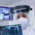
Learn why dental X-rays are essential for accurate diagnosis and treatment planning.
Diagnostic imaging plays a crucial role in dentistry, providing valuable insights into oral health conditions that cannot be easily detected through visual examination alone. By utilizing advanced imaging techniques, dentists are able to assess and diagnose a wide range of oral health issues, from cavities and periodontal disease to jaw alignment and impacted teeth. This allows for more accurate treatment planning and improved patient outcomes.
One of the key advantages of diagnostic imaging in dentistry is its ability to aid in the early detection of dental problems. Dental X-rays, for example, can reveal hidden cavities, bone loss, and infections that may not be apparent during a routine dental checkup. By identifying these issues early on, dentists can intervene promptly, preventing further damage and potentially more extensive and invasive treatment in the future. Additionally, diagnostic imaging can be invaluable in evaluating the alignment of the jaw and bite, informing orthodontic treatment plans, and ensuring optimal results for patients seeking orthodontic correction. Overall, the role of diagnostic imaging in dentistry cannot be overstated, as it enables dentists to provide comprehensive and effective care to their patients.
Dental X-rays are an integral part of comprehensive oral healthcare, playing a crucial role in diagnosing and treating dental conditions. By providing a detailed image of the teeth, jawbone, and surrounding tissues, X-rays offer invaluable insights that aid in identifying hidden dental problems that may not be visible to the naked eye.
One of the primary reasons for the necessity of dental X-rays is their ability to detect dental issues at an early stage. With the use of X-rays, dentists can identify cavities and decay that may not have progressed enough to cause symptoms. This early detection enables prompt intervention, preventing the need for more extensive and invasive treatments later on. Additionally, X-rays help dentists evaluate the development of teeth in children, ensuring proper alignment and timely intervention if any problems arise.
Moreover, dental X-rays prove instrumental in assessing the health of the jawbone and supporting structures. They aid in diagnosing periodontal disease, which affects the gum and bone surrounding the teeth, enabling dentists to design effective treatment plans. X-rays also provide valuable information when evaluating jaw and bite alignment, helping dentists determine the need for orthodontic interventions. Overall, understanding the necessity of dental X-rays is crucial in recognizing their remarkable role in maintaining optimal oral health for patients of all ages.
Dental X-rays are an essential diagnostic tool used in dentistry to assess oral health and detect underlying dental problems. The science behind dental X-rays lies in the use of electromagnetic radiation to capture detailed images of the teeth, bones, and surrounding tissues. This non-invasive imaging technique allows dentists to visualize areas that are not visible to the naked eye, enabling them to make accurate diagnoses and develop appropriate treatment plans.
X-rays work by passing a controlled amount of radiation through the oral cavity, which is absorbed by the different tissues at varying levels. Dense structures, such as teeth and bones, absorb more radiation and appear as white areas on the X-ray image, while less dense structures, like soft tissues, absorb less radiation and appear as darker areas. This stark contrast in image allows dentists to identify existing dental conditions, such as cavities, gum disease, or abnormalities in the bone structure.
In addition to assessing oral health, dental X-rays also play a crucial role in detecting potential problems at early stages. This early detection facilitates prompt intervention, preventing the progression of dental issues and minimizing the need for more invasive procedures in the future. The science behind dental X-rays provides dentists with valuable insights into the underlying health of the teeth, allowing for targeted and effective treatment strategies.
Dental X-rays play a crucial role in the early detection of dental problems, helping dentists identify issues that may not be visible during a routine oral examination. By capturing detailed images of the teeth, jaw, and surrounding structures, dental X-rays provide dentists with vital information to assess oral health and diagnose potential problems.
One of the primary benefits of dental X-rays is their ability to detect cavities in their early stages. X-rays can reveal decay between the teeth or beneath existing dental restorations, allowing dentists to intervene before the cavities grow larger and require more invasive treatment. Additionally, dental X-rays enable dentists to identify other dental issues, such as infections, impacted teeth, or abnormalities in the jawbone structure, which may not be visible to the naked eye.
| Dental Problem | How X-rays Aid in Early Detection |
|---|---|
| Cavities | X-rays can reveal cavities between teeth and beneath fillings. |
| Gum Disease | X-rays show bone loss, a sign of periodontal (gum) disease. |
| Impacted Teeth | X-rays can detect impacted teeth, including wisdom teeth. |
| Abscesses | X-rays reveal abscesses or infections at the tooth’s root. |
| Jawbone Issues | X-rays can identify issues like fractures or abnormalities in the jawbone. |
| Tumors or Cysts | X-rays may uncover tumors, cysts, or other abnormal growths. |
| Tooth Alignment | X-rays help assess tooth alignment and identify potential issues. |
| Developmental Problems | X-rays aid in identifying developmental abnormalities in teeth. |
| Dental Trauma | X-rays assist in diagnosing fractures or other trauma to teeth. |
In summary, dental X-rays aid in the early detection of dental problems by providing dentists with a comprehensive view of the teeth and surrounding structures. By identifying issues in their early stages, dentists can develop appropriate treatment plans, prevent further complications, and maintain their patients’ oral health.
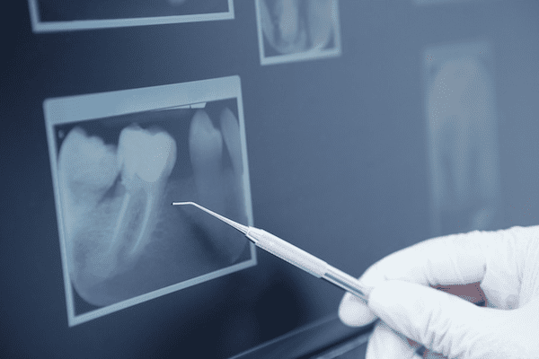
Dental x-rays play an essential role in identifying cavities, providing invaluable information that is often not visible to the naked eye. Cavities, also known as dental caries or tooth decay, are a common oral health issue that affects people of all ages. These small holes in the teeth are caused by the buildup of plaque and bacteria and can lead to further complications if left untreated.
By utilizing dental x-rays, dentists are able to detect cavities in their early stages, allowing for prompt intervention and treatment. This early detection is crucial, as it can prevent the progression of decay and potential damage to the tooth structure. X-rays provide a detailed image of the teeth and their underlying structures, enabling dentists to identify cavities hidden between teeth or beneath existing dental restorations. With this knowledge, dentists can develop personalized treatment plans to address the cavities and restore optimal oral health for their patients.
In addition to their role in early detection, dental x-rays also aid dentists in determining the severity and extent of cavities. By analyzing the x-ray images, dentists can assess the depth of decay and evaluate whether it has reached the inner layers of the teeth, such as the dentin or pulp. This information is vital in determining the appropriate treatment approach, whether it be a simple dental filling or a more extensive procedure such as a root canal therapy.
Overall, the benefits of dental x-rays in identifying cavities cannot be overstated. They provide dentists with critical information to diagnose, treat, and monitor dental caries effectively. Through early detection and accurate assessment, dental x-rays help to preserve the natural teeth and promote long-term oral health for patients.
Dental X-rays play a crucial role in assessing oral health by providing valuable insights that cannot be obtained through visual examination alone. These diagnostic images allow dentists to identify and address potential dental issues at an early stage, preventing further complications and ensuring optimal oral health.
One of the key benefits of dental X-rays in assessing oral health is their ability to detect hidden dental problems. X-rays can reveal tooth decay, cavities, and infections that are not visible to the naked eye. By identifying these issues early on, dentists can devise effective treatment plans to address the problems promptly and prevent them from progressing. Furthermore, dental X-rays aid in the evaluation of bone health and the detection of periodontal disease, allowing for targeted interventions and potential prevention of tooth loss.
| Aspect of Oral Health | Importance of Dental X-rays |
|---|---|
| Detection of Cavities | Dental X-rays can reveal cavities between teeth or beneath fillings, where they might not be visible during a clinical examination. This aids in early intervention and prevents further decay. |
| Assessing Tooth Roots | X-rays help in evaluating the health of tooth roots, detecting issues such as infections, abscesses, or abnormalities in the root structure, which can impact overall oral health. |
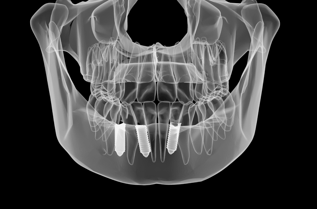
| Monitoring Bone Health | X-rays provide valuable information about the density and condition of the jawbone. This is crucial for assessing bone loss due to periodontal disease and planning treatments like dental implants. |
| Identifying Impacted Teeth | X-rays can reveal impacted teeth (teeth that are unable to erupt properly) such as wisdom teeth, which may cause pain, infection, or damage to adjacent teeth if left untreated. |
| Diagnosing Oral Pathologies | X-rays assist in diagnosing oral diseases and conditions such as cysts, tumors, or developmental abnormalities, which may not be apparent through a visual examination alone. |
| Treatment Planning | Dental X-rays guide dentists in developing comprehensive treatment plans by providing a detailed view of the oral structures, enabling them to tailor treatments according to individual needs. |
In addition to detecting oral diseases and infections, dental X-rays are also instrumental in assessing the alignment of the jaw and bite. These images enable dentists to identify any orthodontic issues, such as malocclusions or misaligned teeth, that may require corrective measures. By utilizing X-rays, dentists can accurately diagnose these problems and develop personalized treatment plans, including orthodontic interventions, to improve bite alignment and overall oral health.
Overall, the importance of dental X-rays in assessing oral health cannot be overstated. These imaging techniques provide dentists with invaluable information about hidden dental problems, bone health, periodontal disease, and bite alignment. By utilizing X-rays as part of regular dental check-ups, dentists can detect problems early on, intervene promptly, and ensure optimal oral health for their patients.
Dental X-rays play a crucial role in assisting dentists in the diagnosis of periodontal disease. Periodontal disease, also known as gum disease, is a common oral health condition that affects the supporting structures of the teeth, including the gums, bone, and periodontal ligaments. While some signs and symptoms of periodontal disease can be observed during a dental examination, X-rays provide valuable information that may not be visible to the naked eye.
One of the key benefits of dental X-rays in diagnosing periodontal disease is their ability to reveal the extent of bone loss caused by the disease. X-rays allow dentists to assess the density and condition of the underlying bone, which is essential in determining the stage of the disease and planning a suitable treatment approach. Additionally, X-rays can help identify any pockets or gaps between the teeth and gums, which are common indicators of periodontal disease. By detecting these issues at an early stage through X-rays, dentists can intervene promptly and prevent further progression of the disease, thus promoting better oral health outcomes for their patients.
Dental X-rays play a crucial role in the evaluation of jaw and bite alignment. By capturing images of the teeth and surrounding structures, these diagnostic images provide important information that aids in diagnosing and planning treatment for patients with alignment issues.
One of the main benefits of dental X-rays in evaluating jaw and bite alignment is their ability to detect any underlying orthodontic problems. X-rays can reveal the position and alignment of both the upper and lower jaws, helping dentists to identify issues such as malocclusions, overbites, underbites, and crossbites. This detailed imagery allows for a more accurate diagnosis and helps dentists develop appropriate treatment plans tailored to the individual patient’s needs. Whether it’s orthodontic treatment or surgical interventions, dental X-rays provide valuable insights that lead to effective and successful outcomes.
Additionally, dental X-rays enable dentists to assess the health and condition of the jawbone and surrounding structures. This is particularly important in cases where patients may have experienced trauma or have missing teeth that can affect their bite. X-rays can reveal any changes in bone density, detect any abnormalities in the jaw joint (temporomandibular joint), and uncover the presence of cysts or tumors that may be impacting the alignment of the jaw. By evaluating these factors, dentists can determine the best course of action to restore proper jaw and bite alignment, leading to improved oral health and overall well-being.
Orthodontic treatment plays a crucial role in correcting misaligned teeth and improving overall oral health. However, determining the most effective treatment plan requires a comprehensive assessment of the patient’s dental structure. This is where dental X-rays become essential.
Dental X-rays provide orthodontists with valuable insights into the patient’s current dental condition. By capturing detailed images of the teeth, jawbone, and surrounding structures, X-rays allow orthodontists to evaluate the alignment of teeth, the positioning of roots, and the presence of any abnormalities. These images help in planning the appropriate orthodontic treatment, whether it involves braces, aligners, or other interventions. Furthermore, dental X-rays enable orthodontists to monitor the progress of the treatment as it unfolds, ensuring that the desired outcome is being achieved and making any necessary adjustments along the way. With the invaluable information obtained from dental X-rays, orthodontists can create personalized treatment plans that lead to beautiful smiles and improved oral health for their patients.
Dental X-rays play a crucial role in identifying impacted teeth and aiding in their treatment. Impacted teeth occur when a tooth fails to erupt fully or remains trapped beneath the gum line. This can occur due to a lack of space in the mouth, misalignment of teeth, or abnormal growth patterns.
Dental X-rays provide vital information about impacted teeth by revealing their location, orientation, and depth within the jawbone. With this valuable insight, dentists can accurately diagnose the condition and determine the most appropriate treatment plan. By identifying impacted teeth early on, dental X-rays help prevent potential complications such as crowding, infection, and damage to neighboring teeth. With the aid of dental X-rays, dentists can effectively plan and execute procedures like orthodontic interventions, extractions, or surgical removals, ensuring optimal oral health outcomes for patients.

Assessing bone loss and infections is a critical aspect of dental care, and dental x-rays play a significant role in this process. These imaging techniques provide valuable insights into the health of the jawbone, helping dentists identify and evaluate conditions such as periodontal disease and various infections. By detecting bone loss at an early stage, dental x-rays allow for proactive treatment planning and the prevention of further damage.
In addition to assessing bone loss, dental x-rays are crucial in identifying and diagnosing infections in the oral cavity. These infections can range from relatively common conditions such as dental abscesses to more severe cases like osteomyelitis, which can lead to significant complications if left untreated. Dental x-rays enable dentists to visualize the extent and location of infections, aiding in accurate diagnosis and the formulation of appropriate treatment strategies. Moreover, they provide a means for monitoring the progress of treatment and ensuring its effectiveness.
Overall, the significance of dental x-rays in assessing bone loss and infections cannot be overstated. Through their ability to provide detailed and accurate images of the jawbone and oral structures, these imaging techniques empower dentists to make informed decisions and provide optimal care for their patients. By utilizing the valuable information provided by dental x-rays, dentists can effectively manage bone loss, prevent complications, and promote oral health.
Dental X-rays play a crucial role in the early detection of tumors and cysts within the oral cavity. These imaging techniques provide detailed images of the teeth, jaws, and surrounding structures, allowing dentists to identify abnormal growths that may be indicative of serious conditions. By using X-rays, dentists can assess the size, location, and extent of tumors and cysts, aiding in their accurate diagnosis and subsequent treatment planning.
Tumors and cysts in the oral cavity can range from benign growths to cancerous lesions. Dental X-rays enable dentists to visualize these abnormalities, even if they are not yet causing noticeable symptoms. Detecting tumors and cysts at an early stage is essential for timely intervention and improved treatment outcomes. With the help of precise imaging techniques provided by dental X-rays, dentists can promptly refer patients to specialists, such as oral surgeons or oncologists, for further evaluation and management. By incorporating dental X-rays as a routine part of oral examinations, dentists contribute to the overall health and well-being of their patients.
Dental X-ray procedures are an integral part of diagnosing and treating various oral health conditions. These procedures require utmost care and safety measures to ensure the well-being of both the patient and the dental professional. The American Dental Association (ADA) provides clear guidelines on the safety measures that should be followed during dental X-ray procedures, prioritizing the health and safety of patients.
First and foremost, dental professionals prioritize the use of lead aprons and thyroid collars to minimize radiation exposure. These protective shields are designed to absorb the majority of radiation, reducing the chances of any potential harm. Additionally, lead-backed barriers are used to protect other parts of the patient’s body that are not being imaged. By diligently adhering to these safety measures, dental professionals can confidently perform X-ray procedures, minimizing risks and ensuring patient safety.
Regular dental x-rays are an essential part of maintaining optimal oral health. These imaging techniques allow dentists to detect potential dental issues early on, leading to prompt and appropriate treatment. However, many patients wonder how often they should get dental x-rays. Understanding the recommendations for the frequency of dental x-rays can help patients make informed decisions about their oral healthcare.
The frequency of dental x-rays varies depending on individual factors such as age, oral health history, and risk of dental conditions. According to the American Dental Association (ADA), adults who have a higher risk of dental problems may require dental x-rays more frequently than those with a low risk. For children and adolescents, the ADA advises considering the child’s individual oral health needs and development stage when determining the frequency of dental x-rays. It’s important to note that dental x-rays are typically considered safe, with low levels of radiation exposure. Dentists follow strict guidelines to minimize radiation exposure and ensure patient safety during these procedures. By adhering to the recommended frequency of dental x-rays, patients can effectively monitor their oral health and address any potential issues before they worsen.
With the rapid advancements in technology, the future of dental imaging holds great promise for the field of dentistry. These advancements are revolutionizing the way dental professionals diagnose and treat oral health issues, enabling them to provide more accurate and efficient care to their patients.
One of the key advancements in dental imaging technology is the introduction of cone beam computed tomography (CBCT). CBCT allows for three-dimensional imaging, providing dentists with a more detailed and comprehensive view of the oral structures. This technology is particularly useful in orthodontics and implantology, as it allows for precise planning and placement of orthodontic appliances and dental implants. CBCT also reduces the need for multiple scans, resulting in less radiation exposure for patients.
Another exciting development in dental imaging is the integration of artificial intelligence (AI) algorithms. AI algorithms can analyze dental images and help detect early signs of dental problems, such as cavities and periodontal disease, with greater accuracy. By assisting dentists in the diagnosis process, AI technology can potentially improve patient outcomes and reduce the need for invasive procedures.
In conclusion, the future of dental imaging is bright, with advancements such as cone beam computed tomography and artificial intelligence revolutionizing the field. These advancements have the potential to enhance patient care by providing more accurate diagnoses and treatment planning. As technology continues to evolve, we can expect even more innovative solutions to emerge, further improving the way dentists assess and address oral health issues.
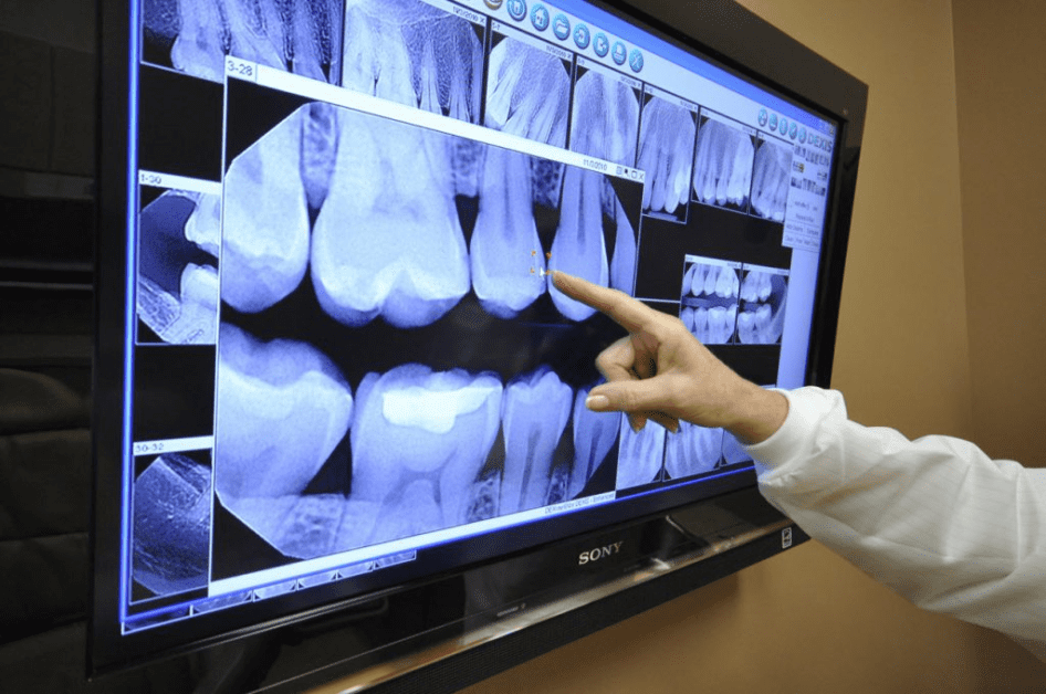
Some advancements in dental imaging technology include digital radiography, cone beam computed tomography (CBCT), and intraoral scanners.
Digital radiography eliminates the need for film development, providing instant high-quality images that can be easily stored and shared electronically.
CBCT is a technology that produces 3D images of the teeth, jaw, and surrounding structures, allowing for more accurate diagnosis and treatment planning.
Intraoral scanners capture detailed 3D images of the teeth and gums, eliminating the need for messy impressions and providing a more comfortable patient experience.
Yes, dental X-rays can aid in the detection of tumors and cysts in the oral cavity, including early signs of oral cancer.
Yes, dental X-rays are considered safe. Dentists take necessary precautions, such as using lead aprons and collars, to minimize radiation exposure.
The frequency of dental X-rays depends on individual patient needs and risk factors. Dentists generally follow the American Dental Association’s guidelines for X-ray frequency.
Yes, dental imaging technology is expected to continue advancing. Researchers are constantly developing new techniques and devices to improve diagnostic capabilities in dentistry.