Physical Address
304 North Cardinal St.
Dorchester Center, MA 02124
Physical Address
304 North Cardinal St.
Dorchester Center, MA 02124

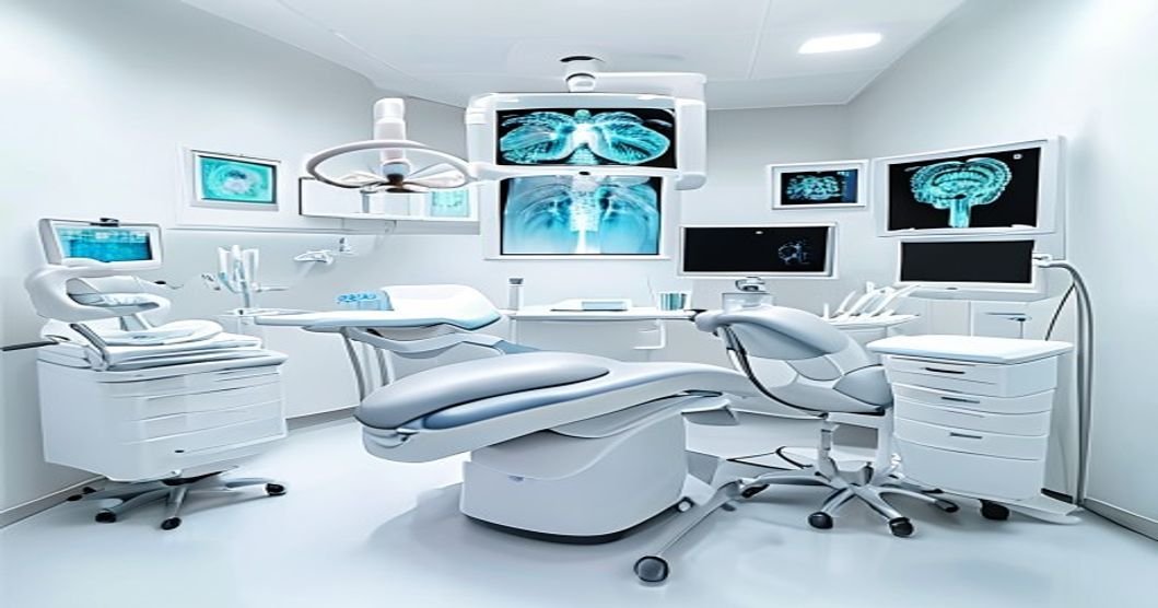
Discover how dental x-rays and imaging protect your oral health. Learn about costs, safety, insurance coverage, and what to expect in our 2026 guide.
When you visit the dentist, you likely expect a visual check of your teeth using a small mirror. However, a significant portion of your oral health exists completely below the surface. Dental X-Rays and Imaging are essential diagnostic tools that allow your dentist to see what the naked eye cannot. These advanced technologies capture detailed pictures of your teeth, roots, jawbone, and surrounding soft tissues. Both children and adults require these routine scans to maintain optimal oral health. Dentists use them to detect hidden cavities, monitor tooth development in children, and assess bone loss in adults. Reading this comprehensive guide will give you a complete understanding of Dental X-Rays and Imaging. You will learn exactly how the procedure works, what it costs, and whether your dental insurance covers it. We will also explore the clinical benefits, potential radiation risks, and what to expect during your appointment. By the end, you will know exactly how this vital technology safeguards your smile and prevents costly dental emergencies.
| Topic | Key Point |
|---|---|
| Primary Purpose | Dental X-Rays and Imaging reveal hidden decay, bone loss, and root issues invisible to the naked eye. |
| Ideal Candidates | All patients, including children, adults, and those planning major dental work like implants or extractions. |
| Procedure Time | The process is incredibly fast, typically taking only 2 to 5 minutes for a standard set of digital images. |
| Frequency | Most patients need bitewing X-rays every 12 to 24 months, while high-risk patients may need them every 6 months. |
| Average Cost | Without insurance, expect to pay between $25 and $250, depending on the type and number of images taken. |
| Insurance Coverage | Most plans cover 80% to 100% of diagnostic imaging costs, often with no waiting period. |
| Safety Profile | Modern digital X-rays emit extremely low radiation, making them exceptionally safe for routine use. |
| Recovery Time | There is zero physical recovery time, allowing you to resume normal activities immediately. |
Dental X-Rays and Imaging involve the use of controlled, low-level electromagnetic radiation to create detailed visual representations of your oral structures. Unlike a standard mirror exam, which only reveals the visible crowns of your teeth, these scans penetrate soft tissues to show the roots, jawbone, and hidden spaces between teeth. To understand why this is necessary, consider basic dental anatomy. A tooth consists of the visible crown and the hidden root, which anchors deep into the jawbone. Decay often begins in the tight contact points between teeth or deep within the grooves. By the time a cavity is visible to the naked eye, it has often progressed significantly into the softer dentin layer.Dental X-Rays and Imaging fit perfectly into a comprehensive preventive oral health care routine. Dentists use various types of imaging depending on the specific clinical need. Bitewing X-rays show the upper and lower back teeth biting together, making them ideal for detecting interproximal decay between teeth. Periapical X-rays capture the entire tooth, from crown to root tip, helping diagnose abscesses, cysts, or root fractures. Panoramic X-rays provide a broad, two-dimensional view of the entire mouth, including the jaw joints, sinuses, and wisdom teeth.Furthermore, advanced 3D imaging, known as Cone Beam Computed Tomography (CBCT), provides a three-dimensional view of the teeth, soft tissues, nerve pathways, and bone density. This is crucial for complex procedures like dental implants, root canals, or wisdom tooth extractions. Together, these tools allow dentists to make accurate, evidence-based diagnoses and formulate effective treatment plans before minor issues become major emergencies.
You might feel perfectly fine, but certain conditions indicate you need a closer look beneath the surface. Here are the most common signs and scenarios that require Dental X-Rays and Imaging.
If you are visiting a new dental practice, a comprehensive set of X-rays is standard protocol. This establishes a baseline of your oral health, revealing any hidden issues that your previous dentist may have been monitoring or treating.
If you experience sharp, lingering pain when biting down or sudden sensitivity to hot and cold temperatures, an X-ray is necessary. These symptoms often indicate decay that has reached the nerve, a cracked tooth, or an infection at the root tip that is not visible above the gumline.
Pimples on the gums, persistent swelling in the jaw, or gums that bleed easily can be signs of an underlying abscess or severe bone loss. Dental X-Rays and Imaging are required to determine the exact source of the infection and the extent of the bone damage.
Before undergoing orthodontic treatment, dental implants, or wisdom tooth removal, detailed imaging is mandatory. The dentist must assess bone density, root angles, and the proximity of nerves to ensure the procedure is safe and predictable.
If you are due for your annual checkup, X-rays will be taken routinely. This does not require an emergency visit. However, if you experience severe, throbbing tooth pain, facial swelling, or trauma to the mouth, call your dentist promptly. Emergency X-rays are needed immediately to rule out fractures or acute infections.
Getting Dental X-Rays and Imaging is a straightforward, non-invasive process. There is no drilling, and absolutely no numbing or anesthesia is required. Here is exactly what happens from start to finish.
Like any medical procedure, it is important to weigh the advantages against the potential drawbacks. Fortunately, modern Dental X-Rays and Imaging have an overwhelmingly positive safety and efficacy profile.
The cost of Dental X-Rays and Imaging is highly affordable, especially when compared to the cost of treating undiagnosed dental disease. Below is a breakdown of typical out-of-pocket expenses in the United States for 2026.
| Item / Service | Average Cost (no insurance) | Typical Insurance Coverage | Notes |
|---|---|---|---|
| Comprehensive Exam | $75 – $150 | 80% – 100% | Required to correlate X-ray findings with clinical exam. |
| Bitewing X-Rays (4 films) | $60 – $120 | 80% – 100% | Standard routine checkup imaging for back teeth. |
| Periapical X-Ray (single) | $25 – $50 | 80% – 100% | Targeted image of a specific painful tooth and root. |
| Full Mouth Series (18 films) | $150 – $250 | 80% – 100% | Comprehensive baseline for new patients, done every 3-5 years. |
| Panoramic X-Ray | $100 – $150 | 80% – 100% | Broad view of jaws, sinuses, and wisdom teeth. |
| CBCT 3D Scan | $250 – $600 | 50% – 80% | Advanced 3D imaging for implants or complex extractions. |
| Typical Total (Routine Visit) | $135 – $270 | Varies | Estimated total including exam and 4 bitewing films. |
Several factors cause cost variations across the country. Geography plays a major role; urban centers and coastal cities generally charge more than rural areas. The provider specialty also affects the price; oral surgeons or endodontists may charge more for specialized imaging like CBCT scans. Additionally, the technology used impacts the final bill, as offices with state-of-the-art digital sensors and 3D imaging capabilities may have slightly higher fees than those using older film-based systems. Finally, the complexity of the case, such as the need for multiple targeted images to diagnose a complex root fracture, will increase the overall fee.
Because Dental X-Rays and Imaging are considered essential diagnostic tools, most dental insurance plans offer excellent coverage for them. The specific ADA procedure codes for these services include D0210 (comprehensive series of radiographic images), D0220 (intraoral periapical first radiographic image), and D0330 (panoramic radiographic image). When your dentist submits a claim, they will use these codes to bill your insurance provider.Coverage largely depends on your plan type. PPO (Preferred Provider Organization) and indemnity plans typically cover 80% to 100% of the cost for diagnostic X-rays, classifying them under “preventive” or “diagnostic” services. HMO (Health Maintenance Organization) plans often cover these services fully with a small, fixed copay. Most plans do not have a waiting period for diagnostic imaging, meaning you can use this benefit immediately upon enrollment. However, insurance companies do enforce frequency limitations. For example, they may only pay for a full mouth series once every 36 to 60 months, and bitewing X-rays only once every 6 to 12 months.If you are unsure about your coverage, ask your dental office to submit a predetermination of benefits. This is a request sent to your insurance company before the procedure to confirm exactly what they will pay and what your out-of-pocket cost will be.If you do not have dental insurance, there are still affordable options. Dental schools often provide diagnostic imaging at a fraction of the private practice cost. You can also look into dental discount plans, which are annual membership programs that provide 20% to 50% off standard fees. Finally, most dental offices offer in-house payment plans to help spread the cost.
While Dental X-Rays and Imaging are the gold standard for diagnosing hidden dental issues, there are a few alternative or supplementary tools a dentist might use in specific situations.
This technology uses a safe, bright light shined through the tooth to detect changes in density. It is preferred for pregnant patients or young children where minimizing any radiation is a top priority. It costs about $10 to $20 as an add-on fee. The key tradeoff is that it can only detect decay on the front or side surfaces of teeth, not deep between the roots or in the jawbone.
This is a tiny, pen-sized camera that takes high-definition, color photographs of the inside of your mouth. It is preferred for patient education, allowing you to see cracked teeth or failing fillings on a monitor. It usually costs $0 to $25 as it is often bundled into the exam fee. The tradeoff is that it only shows the surface of the tooth and cannot see beneath the gumline or inside the bone.
This involves the dentist using a mirror and a metal explorer tool to physically probe the teeth for soft spots. It is preferred in very low-risk patients who strictly refuse all imaging. It costs $0 extra. The major tradeoff is that it is highly unreliable; a tooth can look perfectly healthy on the surface while harboring a massive cavity underneath.
In rare cases of severe facial trauma or suspected tumors, a medical-grade MRI or CT scan ordered by a physician might be used. It is preferred for evaluating soft tissue masses or complex jaw fractures. It costs $500 to $3,000+ and is billed to medical insurance. The tradeoff is the exorbitant cost, long appointment times, and lack of fine detail for individual tooth decay compared to dental-specific imaging.
One of the best aspects of Dental X-Rays and Imaging is that there is virtually no physical recovery time. However, following a few simple aftercare guidelines will ensure you get the most out of your diagnostic visit.
There are no physical restrictions after an X-ray. You can eat, drink, and brush your teeth normally. The primary “aftercare” involves reviewing the results with your dentist. Take the time to look at the images on the screen and ask questions about any dark spots or areas of concern your dentist points out.
If your X-rays revealed a problem, such as a cavity or bone loss, your aftercare involves following the recommended treatment plan. Do not delay scheduling the necessary follow-up procedure, such as a filling or deep cleaning. Delaying treatment allows the hidden issue to grow larger and more painful.
To protect your oral health long-term, adhere to the imaging schedule recommended by your dentist. If you are at high risk for decay, this may mean getting bitewing X-rays every six months. Maintaining excellent daily oral hygiene and a low-sugar diet will help ensure that your future X-rays remain clear and cavity-free.
Call your dentist immediately if you experience any of the following after your visit:
Choosing the right dental professional ensures accurate diagnostics and a comfortable experience. Here are the most important factors to consider when selecting a provider.
When you go in for your consultation, ask these specific questions:
The frequency of Dental X-Rays and Imaging depends entirely on your individual oral health and age. For most healthy adults with a low risk of decay, dentists recommend bitewing X-rays every 12 to 24 months. Children, who are still developing teeth and are more prone to cavities, may need them every 6 to 12 months. High-risk adults, such as those with gum disease or a history of frequent cavities, may also need them every 6 months. Your dentist will tailor the schedule to your specific needs.
Yes, Dental X-Rays and Imaging are considered safe during pregnancy when proper precautions are taken. The radiation dose is exceptionally low, and the X-ray beam is focused solely on the mouth. Furthermore, a lead apron with a thyroid collar is always used to shield the abdomen and chest. However, as a standard precaution, many dentists will postpone routine, non-emergency X-rays until after the first trimester or until after the baby is born, unless there is an acute dental emergency.
No, getting Dental X-Rays and Imaging is completely painless. The process does not involve any needles, drilling, or tissue alteration. The only potential discomfort comes from the physical placement of the digital sensor or film packet in the mouth, which may trigger a mild gag reflex in some patients. The dental team is trained to place the sensors as gently and quickly as possible to minimize any discomfort.
Traditional X-rays use chemical film that must be developed in a darkroom, a process that takes several minutes and exposes the patient to higher radiation levels. Digital X-Rays and Imaging use electronic sensors to capture the image instantly. Digital imaging reduces radiation exposure by up to 80%, eliminates the need for toxic developing chemicals, and allows the dentist to enhance, magnify, and colorize the image on a computer screen for better diagnosis.
Yes, you always have the right to refuse any dental procedure, including Dental X-Rays and Imaging. However, dentists strongly advise against this. Refusing X-rays is like asking a mechanic to fix your car without looking under the hood. The dentist cannot see hidden decay, bone loss, or root infections. Many dental practices have a policy that they cannot provide comprehensive treatment or guarantee their work if they are not allowed to take necessary diagnostic images.
The radiation exposure from Dental X-Rays and Imaging is incredibly low. A standard set of four digital bitewing X-rays exposes you to about 0.005 millisieverts (mSv) of radiation. To put that in perspective, you receive about 0.01 mSv of radiation every single day just from natural background sources like the sun, soil, and the food you eat. A cross-country flight exposes you to more radiation than a full set of dental X-rays.
Yes, this is one of the primary purposes of Dental X-Rays and Imaging. Cavities that form in the tight contact points between teeth, known as interproximal decay, are completely invisible to the naked eye and cannot be felt with a dental explorer tool. Bitewing X-rays are specifically designed to show these areas clearly, appearing as dark shadows on the image, allowing the dentist to catch and treat them early.
Dental X-Rays and Imaging are a safe, fast, and indispensable part of modern preventive dentistry. By revealing hidden decay, bone loss, and root issues, they allow your dentist to intervene early, preventing minor problems from escalating into painful, expensive emergencies. The procedure is entirely painless, requires zero recovery time, and is widely covered by dental insurance plans. If it has been over a year since your last set of X-rays, do not wait for tooth pain to signal a problem. Schedule a consultation with a licensed dentist today to ensure your oral health is thoroughly evaluated. Early intervention guided by accurate imaging is always less expensive, less invasive, and more effective than waiting to treat advanced dental disease.