Table of Contents
Significance of Dental X-Rays in Oral Health Assessment
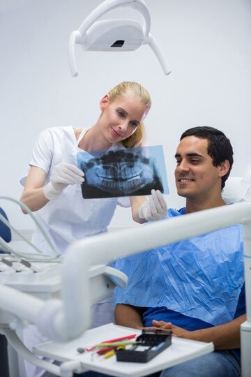
Dental X-rays play a significant role in the assessment of oral health. These diagnostic tools provide valuable insights into the condition of teeth, gums, and surrounding structures that cannot be observed with the naked eye. By capturing detailed images, dental X-rays help dentists identify and diagnose a variety of dental issues, ranging from tooth decay to gum disease. This enables them to develop appropriate treatment plans and formulate preventive measures to maintain optimal oral health.
One of the key benefits of dental X-rays is their ability to detect dental problems in their early stages. By identifying issues such as cavities, infections, and abnormalities early on, dentists can intervene promptly, preventing further damage and potential complications. Dental X-rays are particularly valuable in pediatric dentistry, where children may be unable to communicate problems or exhibit symptoms of dental issues. Through X-ray imaging, dentists can identify underlying problems and address them before they become more severe. This early detection helps ensure that children maintain healthy teeth and gums, promoting their overall oral health as they grow.
Understanding the Purpose of Dental X-Rays
Dental x-rays play a crucial role in understanding and assessing our oral health. They provide invaluable insight into parts of our mouth that are not visible to the naked eye, allowing dentists to accurately diagnose and treat various dental conditions. By capturing detailed images of the teeth, jawbone, and surrounding tissues, dental x-rays enable dentists to detect issues such as tooth decay, bone loss, infections, and even tumors.
One of the primary purposes of dental x-rays is to identify dental problems at their earliest stages. Since many oral health issues may not exhibit visible symptoms initially, x-rays help identify these problems before they become more severe and costly to treat. Dental x-rays also assist dentists in planning and executing specialized procedures such as orthodontic treatments, dental implants, and root canals. With the information obtained from x-rays, dentists can create personalized treatment plans that address the specific needs of each patient.
Exploring the Different Types of Dental X-Rays
Dental X-rays play a crucial role in assessing and diagnosing oral health conditions, and there are several types that dentists may use depending on the specific needs of each patient. One commonly used type is the bitewing X-ray, which provides a detailed view of the upper and lower teeth in a single X-ray image. This allows dentists to evaluate the alignment of the teeth, detect cavities and dental decay, and assess bone health.
Another type of dental X-ray is the periapical X-ray, which focuses on individual teeth and provides a detailed image of the tooth from crown to root. This type of X-ray is useful for identifying issues such as dental abscesses, cysts, or abnormalities in the root canal system. It is particularly beneficial when conducting root canal treatments or evaluating the extent of dental trauma.
Panoramic X-rays are another common type used in dental practices. These X-rays capture a broad view of the entire mouth, including the teeth, jaws, and surrounding structures. Panoramic X-rays are particularly useful for assessing the development of wisdom teeth, identifying jawbone disorders, and detecting tumors or abnormalities in the oral and maxillofacial region.
By utilizing these different types of dental X-rays, dentists are able to gather valuable information about a patient’s oral health. With proper interpretation and analysis, these X-rays contribute to accurate diagnosis, personalized treatment planning, and the maintenance of optimal oral health.
| Type of X-Ray | Description |
|---|---|
| Bitewing X-rays | Used to detect decay between teeth and assess the health of the jawbone. |
| Periapical X-rays | Capture images of the entire tooth, from the crown to beyond the root where it connects to the jawbone. |
| Panoramic X-rays | Provides a broad view of the entire mouth, including all the teeth, upper and lower jaws, and surrounding structures. |
| Occlusal X-rays | Focuses on the bite of the teeth and how the upper and lower teeth fit together when the jaw is closed. |
| Cone Beam Computed Tomography (CBCT) | Offers 3D images of the teeth, jawbone, and surrounding facial structures for detailed assessment. |
Frequency of Dental X-Rays: Determining the Ideal Interval
Determining the ideal interval for dental x-rays can be a complex task, as it depends on various factors such as a patient’s oral health status, age, risk factors, and medical history. The American Dental Association (ADA) recommends that the frequency of dental x-rays should be individualized based on a patient’s specific needs and risk factors.
For adults with good oral health and low risk of dental problems, the interval between dental x-rays can range from 18 to 36 months. However, for patients with a history of gum disease, a higher risk of cavities, or those undergoing orthodontic treatment, more frequent x-rays may be necessary. The ADA guideline suggests that children may require x-rays more frequently than adults due to their changing dentition and higher risk of decay.
The decision regarding the interval of dental x-rays should be made in consultation with a dental professional, who will consider the patient’s individual needs and risk factors. Regular dental check-ups and oral examinations can help dentists assess a patient’s oral health status and determine if dental x-rays are necessary. By tailoring the interval of x-rays to each patient’s specific circumstances, dental professionals can ensure the appropriate balance between diagnostic value and patient safety.
Factors Influencing the Need for Dental X-Rays
Factors influencing the need for dental x-rays are diverse and can vary from patient to patient. One key factor is the individual’s oral health history. For instance, patients with a history of dental problems, such as tooth decay or gum disease, may require x-rays more frequently to monitor their conditions. Additionally, individuals with a greater risk for oral health issues, such as patients with a family history of dental diseases, may also benefit from more frequent x-ray examinations.
Another important factor to consider is the patient’s age and stage of dental development. Pediatric patients, for example, may require x-rays more frequently to monitor the growth and development of their teeth, detect orthodontic problems, and ensure proper alignment. On the other hand, adults may require x-rays less frequently, but routine x-rays are still essential for detecting issues that may not be visible during a regular dental examination, such as cavities between teeth or problems beneath the gumline.
In conclusion, various factors influence the need for dental x-rays. Oral health history, risk factors, age, and stage of dental development all play a vital role in determining the frequency and necessity of x-ray examinations. Dentists must carefully evaluate each patient’s unique circumstances and consider these factors in order to provide the most accurate and comprehensive oral health assessment possible.

The Role of Dental X-Rays in Early Detection of Oral Health Issues
Dental X-rays play a crucial role in the early detection of oral health issues. While a visual examination can provide valuable information, it is limited to what is visible on the surface. Dental X-rays, on the other hand, allow dentists to see what lies beneath, giving them a more comprehensive understanding of a patient’s oral health.
One of the main benefits of dental X-rays is their ability to detect problems that may not be immediately apparent. For example, X-rays can reveal hidden cavities between teeth or beneath fillings, as well as infections in the root canal or surrounding bone. This early detection enables dentists to address these issues promptly before they worsen and potentially become more complicated and expensive to treat.
Additionally, dental X-rays are invaluable when it comes to screening for developmental issues and abnormalities. With X-rays, dentists can identify problems such as impacted teeth, tumors, cysts, or bone abnormalities that might otherwise go unnoticed. By catching these issues early on, dentists can formulate appropriate treatment plans or refer patients to specialists, ensuring optimal oral health outcomes.
Overall, the role of dental X-rays in early detection of oral health issues cannot be overstated. These diagnostic tools provide dentists with vital information that aids in the timely and accurate diagnosis of dental problems. By detecting and addressing issues early, dental X-rays contribute to better oral health outcomes for patients. So, next time you visit your dentist, don’t hesitate to discuss the importance of dental X-rays in your oral health assessment.
Benefits of Regular Dental X-Rays for Preventive Care
Regular dental x-rays play a crucial role in preventive care by providing valuable insights into the oral health of patients. They allow dentists to detect potential issues at their earliest stages, preventing them from progressing into more serious conditions. By capturing detailed images of the teeth and supporting structures, dental x-rays enable dentists to identify cavities, gum disease, developmental abnormalities, and other problems that may not be visible during a routine examination.
Detecting dental problems early through regular x-rays allows dentists to intervene with appropriate treatments before they worsen. For example, dental x-rays can reveal hidden cavities between teeth, preventing the need for more invasive procedures such as root canals. Additionally, x-rays can detect infections or oral diseases that may not show obvious symptoms, allowing prompt treatment and preventing further complications. By addressing oral health issues in their early stages, dental x-rays contribute significantly to maintaining optimal oral health and preventing extensive dental work in the future.
| Benefit | Description |
|---|---|
| Early Detection of Dental Issues | X-rays can detect cavities, gum disease, and other dental problems in their early stages before they worsen. |
| Monitoring Growth and Development | X-rays help monitor the growth and development of teeth and jaws, especially in children and adolescents. |
| Diagnosis of Hidden Dental Problems | X-rays can reveal hidden dental issues such as impacted teeth, abscesses, and tumors that are not visible to the naked eye. |
| Treatment Planning | X-rays aid in the development of treatment plans for various dental procedures, including orthodontic treatments and implants. |
| Tracking Changes Over Time | Regular X-rays allow dentists to track changes in the mouth over time and adjust treatment plans accordingly. |
| Prevention of Serious Complications | Early detection through X-rays can prevent serious complications and costly treatments in the future. |
| Patient Education | X-rays provide visual evidence of dental issues, helping dentists educate patients about their oral health and the need for preventive measures. |
Minimizing Radiation Exposure during Dental X-Rays
When it comes to dental x-rays, one of the main concerns for both patients and healthcare providers is radiation exposure. While dental x-rays are generally considered safe, it is still important to minimize radiation exposure as much as possible. Fortunately, advancements in technology and guidelines have made it possible to reduce radiation exposure during dental x-rays.
One of the key ways to minimize radiation exposure is by using digital x-ray systems. Unlike traditional film x-rays that require higher levels of radiation, digital x-ray systems use a lower dose of radiation to produce high-quality images. These systems also allow for immediate image viewing, eliminating the need for retakes and further exposure. Additionally, digital x-rays can be easily stored and electronically transmitted, reducing the need for physical storage space and unnecessary handling of films.
Another approach to minimizing radiation exposure is by using lead aprons and thyroid collars. These protective accessories are designed to shield the body from unnecessary radiation. Lead aprons are worn by patients during dental x-rays to protect vital organs and tissues, while thyroid collars specifically shield the thyroid gland from radiation. By incorporating these protective measures, the risk of radiation exposure can be significantly reduced.
It is worth noting that while minimizing radiation exposure is important, it should not compromise the diagnostic value of dental x-rays. Dentists carefully assess the individual needs of each patient and determine the appropriate frequency and type of x-rays required for accurate diagnosis and treatment planning. By striking the right balance between minimizing radiation exposure and ensuring effective oral health assessment, dental professionals can provide safe and comprehensive care to their patients.
Alternative Diagnostic Tools to Dental X-Rays
When it comes to diagnosing dental issues, X-rays have long been the go-to tool for dentists. However, advancements in technology have introduced alternative diagnostic tools that can complement or even replace dental X-rays in certain cases. These tools, although not as widely used as X-rays, offer several benefits in terms of minimizing radiation exposure and providing additional diagnostic information.
One such alternative is the use of intraoral cameras. These small cameras are designed to capture detailed images of the teeth and oral cavity. Dentists can use these images to closely examine hard-to-reach areas, identify potential problems, and educate patients about their oral health. Intraoral cameras also allow for real-time visualization, providing immediate feedback and enhancing communication between dentist and patient.
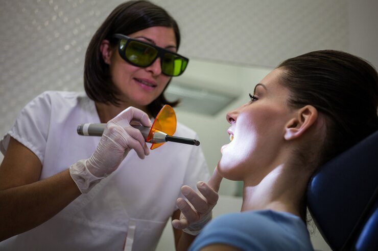
Another tool gaining popularity is dental lasers. These high-tech devices emit a concentrated beam of light that can be used for a variety of procedures, including diagnostics. Dental lasers can help detect cavities and other dental abnormalities by measuring the reflection or fluorescence of light in the oral cavity. This non-invasive technique is particularly useful for detecting early-stage dental caries, as it can identify demineralization that may not be visible to the naked eye.
While these alternative diagnostic tools offer promising advantages, it’s important to note that they are not meant to completely replace dental X-rays. X-rays remain an essential tool for comprehensive oral health assessment, especially when it comes to evaluating the condition of the underlying bone and detecting certain dental issues such as abscesses or impacted teeth. Dentists carefully consider each patient’s unique needs before determining which diagnostic tools, including alternatives to X-rays, are best suited for their particular case.
Addressing Common Misconceptions about Dental X-Rays
Dental x-rays have long been a subject of concern and misconceptions among patients. One of the most common misconceptions is that dental x-rays are unnecessary and only expose patients to harmful radiation. However, it is important to understand that dental x-rays play a crucial role in assessing oral health and detecting potential issues that may not be visible during a routine dental examination.
Contrary to popular belief, dental x-rays emit very low levels of radiation and are considered safe for patients of all ages. In fact, the amount of radiation received during a dental x-ray is equivalent to the radiation exposure experienced during a short airplane flight. Dental professionals take extensive precautions to minimize radiation exposure, such as using lead aprons and thyroid collars to protect other parts of the body.
Moreover, dental x-rays are an essential tool for diagnosis and treatment planning. They allow dentists to identify cavities, bone loss, impacted teeth, and other abnormalities that may not be visible to the naked eye. This helps in early detection and prevention of oral health issues, ultimately saving patients from potential pain, discomfort, and costly treatments in the long run.
It is important to note that the frequency of dental x-rays is determined based on each individual’s oral health needs. Dentists carefully consider factors such as age, dental history, presence of symptoms, and risk factors before recommending x-rays. By addressing these misconceptions and providing accurate information, patients can make informed decisions about their oral health and understand the importance of dental x-rays in maintaining a healthy smile.

Collaboration between Dentists and Patients in X-Ray Decision-Making
Collaboration between dentists and patients in X-ray decision-making is crucial for ensuring the appropriate and necessary use of this diagnostic tool. Dentists have the expertise and knowledge to determine when X-rays are required based on a thorough assessment of a patient’s oral health. However, patients also play an important role in this process by providing valuable information about their medical history and any specific concerns they may have.
Open communication between dentists and patients is key in making informed decisions regarding the need for dental X-rays. Dentists should take the time to explain the purpose of the X-rays, the potential benefits, and any associated risks or alternatives. Patients, on the other hand, should feel comfortable asking questions and expressing any concerns they may have. This collaborative approach helps foster trust and ensures that patients are active participants in their own healthcare decisions.
By working together, dentists and patients can make informed and personalized decisions regarding the use of dental X-rays. Dentists can leverage their expertise to recommend the appropriate type and frequency of X-rays based on individual patient needs, while patients can provide essential input to ensure that their concerns and preferences are taken into account. This collaborative process ultimately promotes patient-centered care and enhances the overall oral health outcomes for individuals.
Evaluating the Cost-Effectiveness of Dental X-Rays
Dental x-rays are a valuable tool in the field of dentistry, allowing dentists to gain a deeper understanding of their patients’ oral health. However, it is essential to evaluate their cost-effectiveness to ensure that patients are receiving the best care possible while minimizing unnecessary expenses.
When assessing the cost-effectiveness of dental x-rays, factors such as the specific needs of the patient, the type of x-ray being performed, and the potential benefits derived from the procedure should be considered. For instance, in cases where a patient presents with symptoms of tooth decay or gum disease, dental x-rays can provide detailed images of the affected areas, helping dentists make accurate diagnoses and develop appropriate treatment plans.
Furthermore, dental x-rays are instrumental in detecting issues that are not visible to the naked eye, such as impacted teeth, abscesses, or jawbone infections. By identifying these problems early on, dentists can intervene promptly, preventing further complications and potentially saving patients from extensive and costly procedures in the future.
While it is crucial to evaluate the cost-effectiveness of dental x-rays, it is equally important to strike a balance between financial considerations and the overall health and well-being of patients. Dentists must carefully assess each individual case, considering the potential benefits and risks associated with dental x-rays, to make informed decisions that prioritize the best interests of their patients.
Conclusion: Balancing the Need for Dental X-Rays with Patient Safety
When it comes to dental x-rays, finding the right balance between the need for diagnostic information and patient safety is crucial. While dental x-rays play a vital role in assessing oral health and detecting potential issues, it is equally important to minimize radiation exposure and ensure the overall well-being of patients.
The American Dental Association (ADA) and the Food and Drug Administration (FDA) have set guidelines to help dentists determine the appropriate use of dental x-rays. These guidelines take into consideration factors such as age, oral health history, and risk of dental problems. Dentists are trained to assess individual patient needs and make informed decisions regarding the frequency and type of x-rays required.
By adhering to these guidelines and using advanced techniques such as digital x-rays and lead aprons, dental professionals can significantly reduce radiation exposure while still providing accurate diagnostic information. It is important for dentists and patients to work together in the decision-making process, weighing the potential benefits of dental x-rays against the risks. By striking the right balance, dental x-rays can be a valuable tool in maintaining oral health and preventing more serious dental issues down the line.
How often should dental X-rays be taken?
The frequency of dental X-rays depends on various factors such as the patient’s oral health, age, and risk of developing dental problems. It is typically recommended to have X-rays taken every 1-2 years for most adults.
Are dental X-rays safe?
Dental X-rays are generally considered safe, as the radiation exposure is minimal. Dentists take precautions to minimize radiation exposure by using lead aprons and collars. Additionally, digital X-ray technology further reduces radiation exposure compared to traditional X-rays.
Can dental X-rays detect oral health issues early?
Yes, dental X-rays play a crucial role in the early detection of oral health issues. They can reveal hidden cavities, bone loss, impacted teeth, infections, and other dental problems that may not be visible during a regular examination.
Are there alternative diagnostic tools to dental X-rays?
While dental X-rays are highly effective, there are alternative diagnostic tools available. In some cases, dentists may use intraoral cameras, 3D imaging, or visual examinations to assess oral health. However, these alternatives may not provide the same level of detail as X-rays.
How can patients minimize radiation exposure during dental X-rays?
Patients can minimize radiation exposure by wearing lead aprons and collars provided by the dentist. Additionally, digital X-rays emit less radiation than traditional X-rays, so opting for digital imaging can further reduce exposure.
What are some common misconceptions about dental X-rays?
One common misconception is that dental X-rays are unnecessary and harmful. However, X-rays are crucial for diagnosing dental problems. Another misconception is that pregnant women should avoid X-rays, but with appropriate shielding, dental X-rays can be safe during pregnancy.
Should patients be involved in the decision-making process for dental X-rays?
Yes, collaboration between dentists and patients is essential in making decisions regarding dental X-rays. Dentists should explain the purpose and benefits of X-rays, while patients can voice their concerns or preferences. Ultimately, both parties should make an informed decision together.
How cost-effective are dental X-rays?
Dental X-rays are generally considered cost-effective as they help detect dental problems early, potentially preventing more extensive and expensive treatments in the future. It is important to weigh the cost of X-rays against the potential benefits and long-term savings in dental care.

